Please enter url.
Login
Logout
Please enter url.
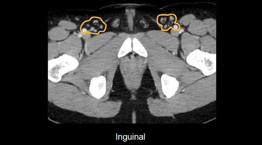
Abdominal CT: lymph nodes • LITFL • Radiology Library
litfl.com
source
Comments
Abdominal CT: lymph nodes • LITFL • Radiology Library
Lipiodol lymphangiography. The injected lipiodol flowed up through the ...
Patient in left lateral decubitus position, with a view of the extent ...
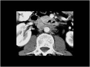
Computed tomography (CT) scan of the abdomen and pelvis demonstrating ...
CT-scan of the second case showing a bladder wall thickening, with ...
Types of variations of ICA. a Straight course, b tortuosity, c kinking ...
CECT Abdomen axial image showing complete luminal occlusion of External ...
Computed tomography image of a schwannoma located in the posterior ...
Article - A simplified approach to the spaces of the suprahyoid neck
Urinary bladder Paraganglioma, A case report | Eurorad
Tracheal Paraganglioma: Differential Diagnosis of a Contrast-Enhanced ...
Diagnosis of sarcopenia using the PMI. Example: A 67-year-old woman ...
Thyroid teratoma – a challenging diagnosis of a thyroid nodule | Eurorad
Mesenteric cyst: a rare cause of abdominal pain | Eurorad
Femoral vein thrombophlebitis and septic pulmonary embolism from ...
Thyroid Cancer Invades the Trachea - Chest Case Studies - CTisus CT ...
Vascular involvement in polyarteritis nodosa | Eurorad
CT scan images of aortic dissection (arrows) extending in the left ...
Resection of portion of large bowel within Indiana pouch diversion ...
Contrasted CT showing para-aortic lymph node bulking (limited by blue ...
Symptomatic radiation recall pneumonitis induced by immunotherapy | Eurorad
Frontiers | Neoadjuvant Multikinase Inhibitor in Patients With Locally ...
LearningRadiology.com-Abdominal Pain
Image | Radiopaedia.org
Urinary Tract and male reproductive system | 2.1 Kidney and ureter ...
Bilateral Inguinal Hernia Containing Urinary Bladder as Sole Content ...
(PDF) Aortoduodenal fistula successfully treated with endovascular repair
Vanishing bone lesions mimicking osteoblastic metastases | Eurorad
Abdomen and retroperitoneum | 1.9 Retroperitoneum and great vessels ...
ER radiology evaluation of appendicitis and alternative diagnoses of ...
Tracheal Paraganglioma: Differential Diagnosis of a Contrast-Enhanced ...
Enlarged diameter of trachea (33.6 mm×37.5 mm) in inspiration computed ...
Abdomen and retroperitoneum | 1.9 Retroperitoneum and great vessels ...
CT and ultrasound. Axial thoracic contrast-enhanced CT images at ...
Piriform sinus carcinoma | Eurorad
Right-Inguinal-Lymph-Node
Inguinal-Lymph-Nodes-Diagram
Axillary-and-Inguinal-Lymph-Nodes
Inguinal-Lymph-Node-Biopsy
Bilateral-Inguinal-Lymph-Nodes
Inguinofemoral-Lymph-Nodes
Pelvic-Lymph-Node-Anatomy
Abdominal-Lymph-Nodes-Anatomy
Inguinal-Nodes-Location
Lymph-Node-Sinuses
Inguinal-Lymph-Node-Dissection-Anatomy
Lymph-Nodes-Gross-Anatomy
Abdomen-Lymph-Node-Anatomy
Popliteal-Nodes
Inguinal-Lymph-Node-Surgery
Groin-Lymph-Node-Swelling