Please enter url.
Login
Logout
Please enter url.
Hashimoto Encephalopathy: Rare Cause of Stroke in Young Pati... : The ...
journals.lww.com
source
Comments
Comparison of myelin oligodendrocyte glycoprotein (MOG)-antibody ...
Brain Resection | MRI Timeline
Degenerative Diseases | Radiology Key
Postpartum Angiopathy With Reversible Posterior Leukoencephalopathy ...
FLAIR and T2*-weighted images of ARIA-E event. (A) FLAIR (top row) and ...
MRI lesions in the acute stage of cryptogenic new-onset refractory ...
Clinical presentations of five patients with VARS2 gene mutation ...
Motor Recovery and Cortical Reorganization After Mirror Therapy in ...
Clinical images of Case 1. (A1) Brain MRI with contrast of brain for ...
Case report with focused review
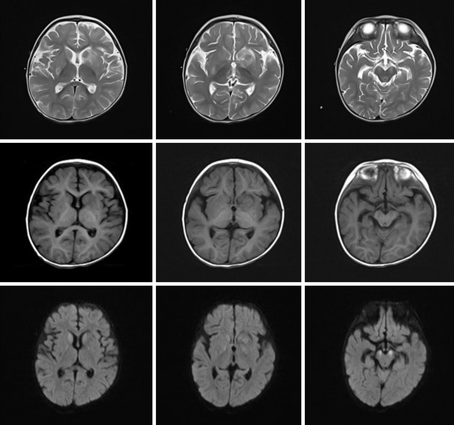
Emergence of lesions outside of the basal ganglia and irreversible ...
Leigh syndrome | pacs
Figure 2 from A Pathology-proven Case of Schilder's Disease | Semantic ...
Figure 1 from [A case of acute hyperammonemic encephalopathy with ...
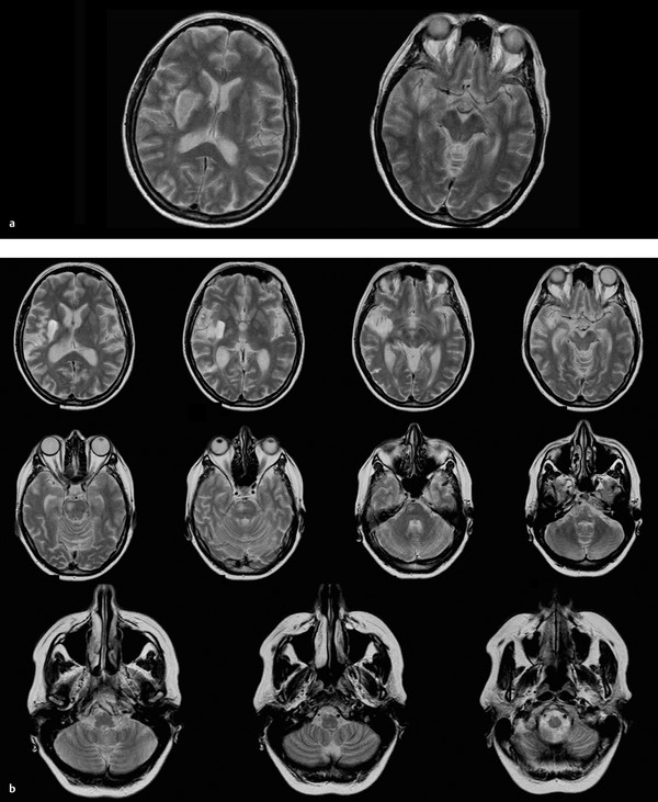
A: Axial MR images of the brain in the axial plane, T2W FLAIR, showing ...
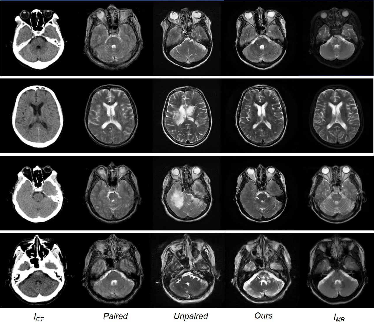
GitHub - ChengBinJin/MRGAN-TensorFlow: This repository is the ...
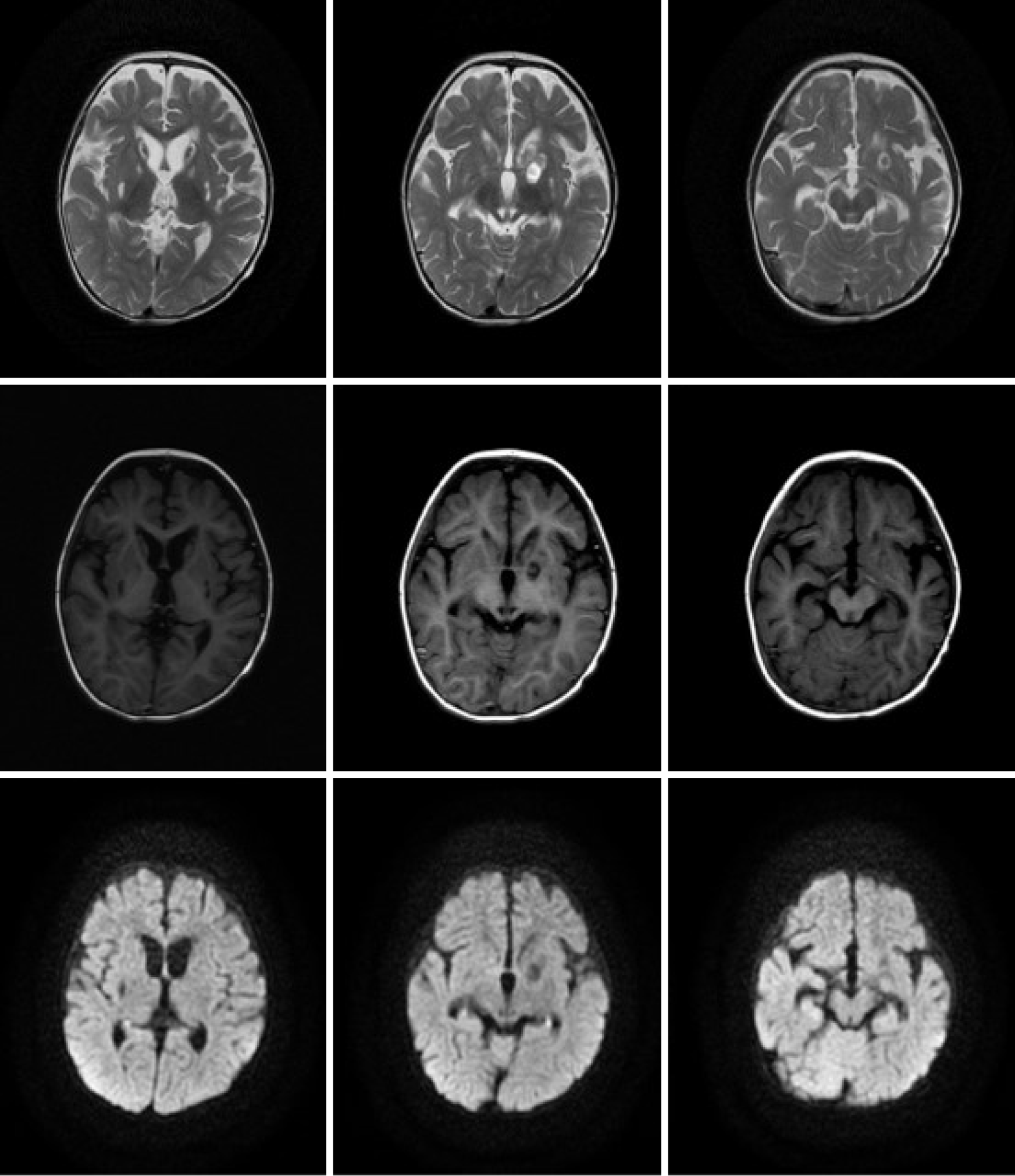
Serial axial T2-weighted images of the brain at the level of the corona ...
Emergence of lesions outside of the basal ganglia and irreversible ...
Tumor-like lesion in NMO. Axial and sagittal planes show the large ...
MRI formation of symmetrical hyperintensities in the putamen and ...
Emergence of lesions outside of the basal ganglia and irreversible ...
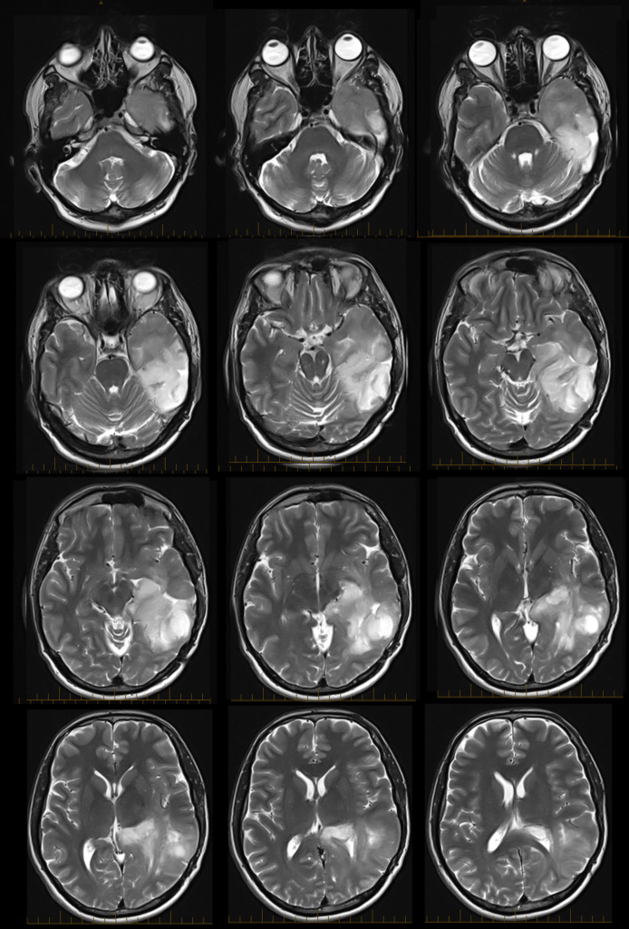
Axial head magnetic resonance and computed tomography scans. a ...
T2-weighted MRI images for patient Y.Q. The right side of each image ...
Figure2.Cranial magnetic resonance imaging of the patient. a: Axial ...
Atypical Diffusion-Restricted Lesion in 5-Fluorouracil Encephalopathy ...
Rosette-forming glioneuronal tumor: an illustrative case and a ...
MRI sequences related to patient’s PRES. Top row Diffusion, Middle and ...
MR Imaging of Human Herpesvirus-6 Encephalopathy after Hematopoietic ...
Familial Sneddon’s syndrome with microbleeds in MRI | BMJ Case Reports
:: BNR :: Brain & Neurorehabilitation
Diffusion‐weighted magnetic resonance imaging reveals symmetrically ...
Aqueductal tumors — axial view | Download Scientific Diagram
Figure 1 from Autoimmune glial fibrillary acidic protein astrocytopathy ...
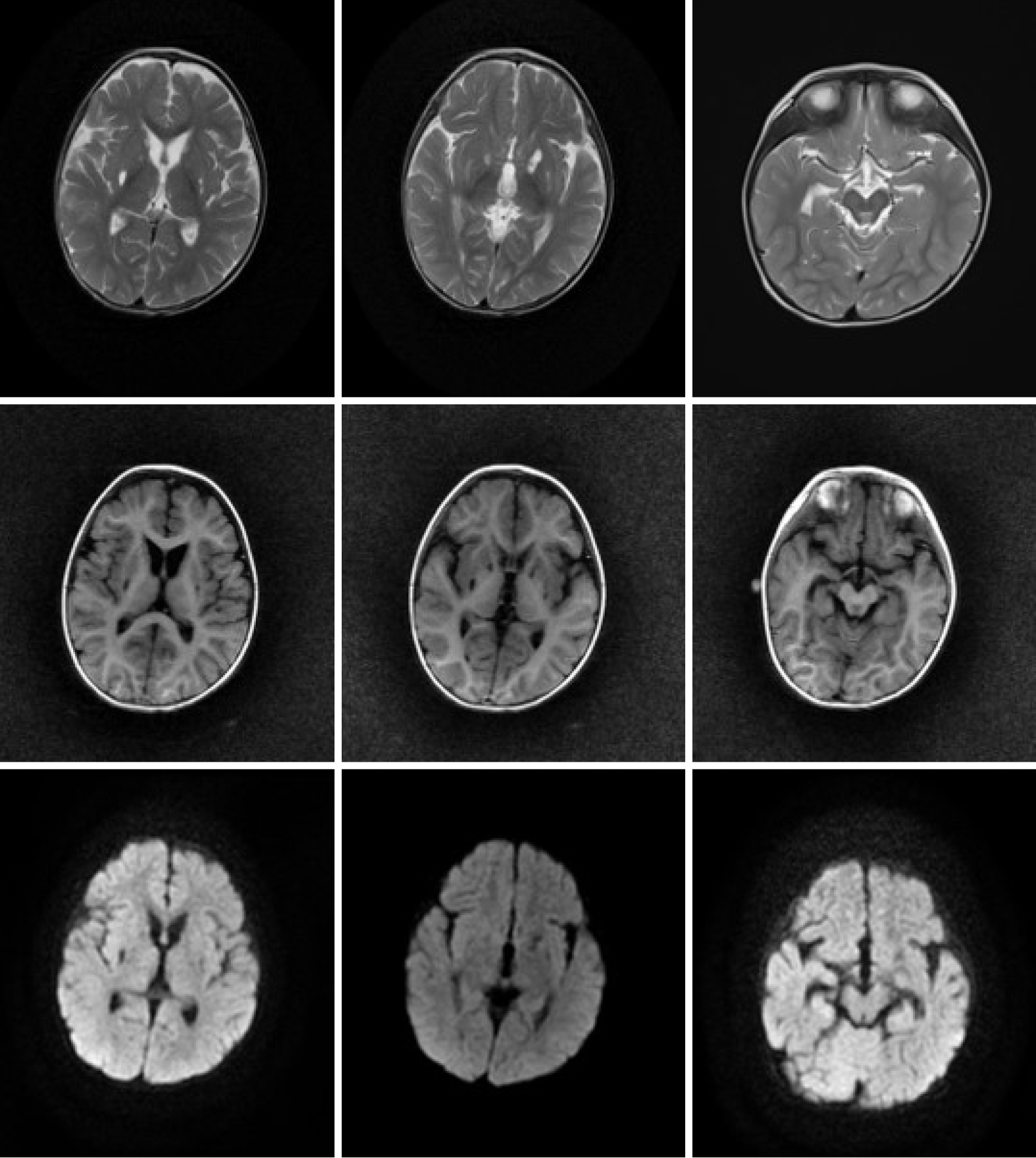
White matter lesions observed through three different sequences of MRI ...