Please enter url.
Login
Logout
Please enter url.
(A–C): Salt and pepper sign. FLAIR axial MRI image of the left temporal ...
researchgate.net
source
Comments
(A, B): Optic nerve tram-track sign. Postcontrast T1W axial MRI image ...
(Case 2) Axial brain MRI indicating a lesion in the right frontal lobe ...
Head injury | Neupsy Key
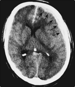
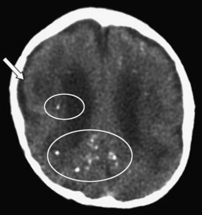
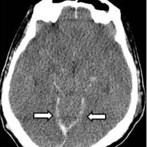
CT scan showed high‐density shadow interhemispheric fissure cistern ...
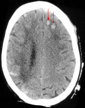
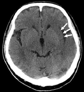
A 68-year-old woman with a left hemiplegia following a conscious ...
A 68-year-old woman with a left hemiplegia following a conscious ...
The soft-tissue-window of a cerebral CT of a young patient demonstrates ...
Brain Abscess Imaging: Practice Essentials, Radiography, Computed ...
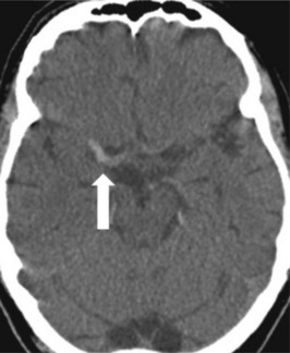
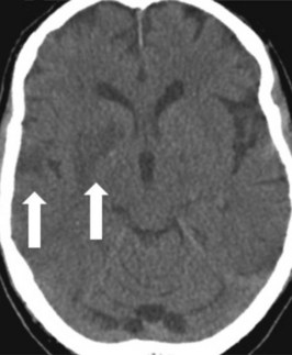
(PDF) Hyperdense middle and anterior cerebral arteries: Familiar and ...
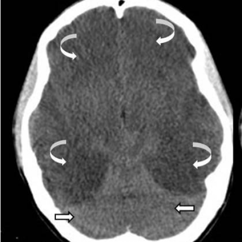
(PDF) Case 14694 Severe hypoxic-ischemic brain injury: the reversal ...
(PDF) Hypoparathyroidism presenting with seizures and intracranial ...
Head CT
Dural Calcifications: Normal Locations and Appearances | SpringerLink
La TC sin contraste realizada a las 48 horas muestra ausencia de ...
Initial CT scan of a 53 year-old man revealing a hyperdense region in ...
Acute encephalopathy of Bacillus cereus mimicking Reye syndrome - Brain ...
Initial brain CT scan: hypodense aspect of the upper brainstem and ...
Sagittal section of brain CT showing significant pneumocephalus with ...
The native CT scan of the brain revealed a subtle oval hypodensity on ...
(A) Scheme showing the bladder and vesicouterine serosal reflection ...
Hyperintense lesions mainly in posterior parieto-occipital area on both ...
(PDF) Neonatal Temporal Lobar Hemorrhage Secondary to Thrombosis of the ...
A 70-year-old woman (case 1). (a, b) Brain CT performed immediately ...
Viral Infections of the Nervous System | Neupsy Key
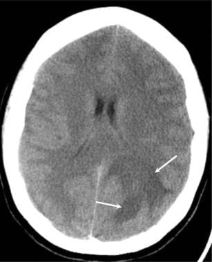
Acute diffuse subdural hemorrhage along the right cerebral hemisphere ...
MRI Brain showing Lesion with Perilesional Edema | Download Scientific ...
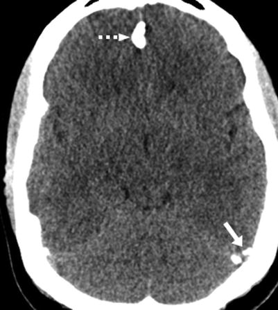
(PDF) Kernohan's Notch: A Forgotten Cause of Hemiplegia—CT Scans Are ...
Axial CT revealing small bilateral isodense subdural hematomas ...
IRM cérébrale Coupes axiales T2 (a,b) ; FLAIR (c,d) ; T1 (e) et T1 ...
Severe hypoxic - ischaemic brain injury: the reversal, the pseudo ...
Diagnosis and Management of Cerebral Venous Thrombosis | Stroke
a CT of the skull after injury reveals hypodense, thin temporal bones ...
Stroke Imaging: Practice Essentials, Computed Tomography, Magnetic ...
A 70-year-old woman (case 1). (a, b) Brain CT performed immediately ...
Severe hypoxic - ischaemic brain injury: the reversal, the pseudo ...