Please enter url.
Login
Logout
Please enter url.
Cervical Spine Mri Labeled
ar.inspiredpencil.com
source
Comments
Normal Cervical Spine MRI Explained | Dr. Jeffrey P.Johnson | HD - YouTube
Evolution of clinically isolated syndrome (CIS) optic neuritis: The ...
...but maybe tomorrow: treatment update and basilar invagination?
Anesthetic management of spontaneous cervical epidural hematoma during ...
Dr Balaji Anvekar FRCR: Basilar Impression (BI)
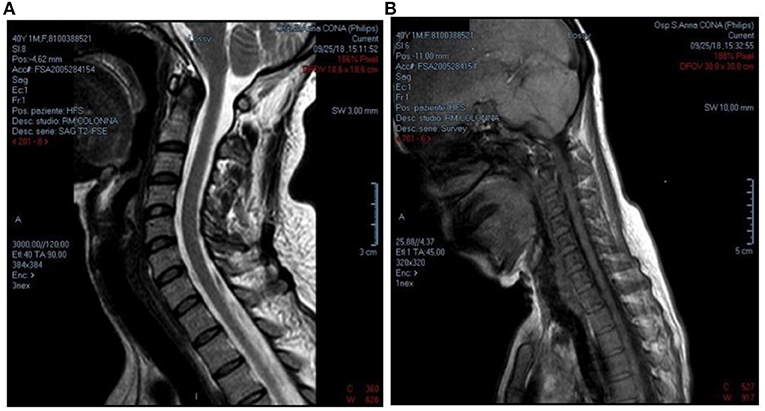
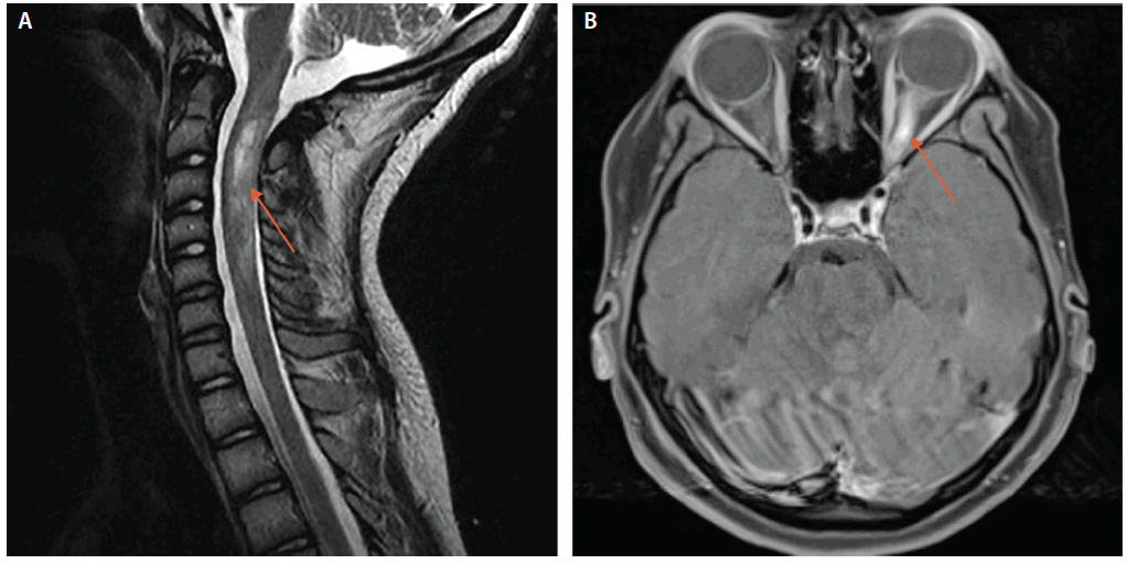
MRI with IV contrast. A: T2-weighted imaging in the sagittal plane with ...
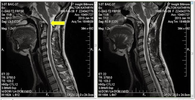
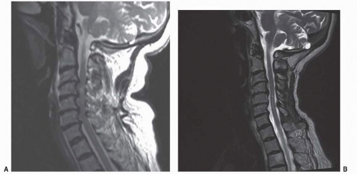
Saggittal T1 cervical spine MRI: day 1 (a) and day 11 (b). Serial MRI ...
(A) Intraoperative fluoroscopy showing closed reduction of the fracture ...
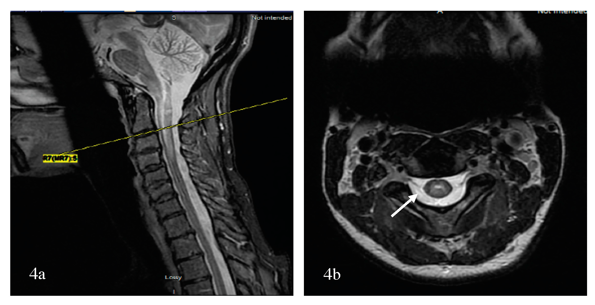
Case 4. a, b T2-weighted sagittal sections of the whole spine showing a ...
The occipitocervical parameters measurement on MRI images. a The ...
Evolution of clinically isolated syndrome (CIS) optic neuritis: The ...
C-5-C-6-stenosis | War in Context
Conservative Care for Cervical & Lumbar Disc Herniations | El Paso Back ...
A: C2-7 sagittal alignment measured as the Cobb angle between the lines ...
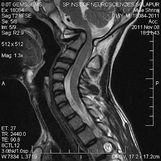
Case 1: MRI of the cervical segment of the spinal cord of a 66-year-old ...
Frontiers | Hirayama Disease: A Case of an Albanian Woman Clinically ...
Brain Sciences | Free Full-Text | Pattern Recognition of the Multiple ...
Follow-up magnetic resonance image demonstrating a syrinx in the ...
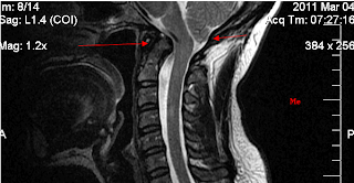
T-2 weighted with fat supression of sagital cervical MRI shows fusiform ...
Neurenteric cyst at the dorsal craniocervical junction in a child: Case ...
C spine flexion | Portland MRI | Siker Medical Imaging
Central Cord Syndrome: Acute Decompression versus Watch and Wait ...
Three-month follow-up T1-weighted brain MRI and T2-weighted spine MRI ...
What is Basilar Invagination? ~ Ramblings of an Incredibly Large Brain
Antibody-Mediated Inflammatory Central Nervous System Disorders of ...
(A) MRI myelogram (B) CT myelogram coronal (C). CT myelogram ...
Slice positioning based on T2-w TSE sagittal images (left ...
Postoperative transient tetraplegia in two patients caused by cervical ...
Multiple spinal neurofibromas with cervical and lumbar cord compression ...
Cervical CT shows spinal cord compression C3–C4 and C4–C5. | Download ...
Traumatic Spondylolisthesis of the Axis without Fracture: Case Report ...
Saggittal T1 cervical spine MRI: day 1 (a) and day 11 ( | Open-i
Sagittal cervical spinal cord T1-weighted MRI demonstrating no ...
The recurrent lingual TGDC in 11-year-old boy in MR imaging. | Download ...
Demyelinating Diseases, Neuro-Oncology, and Disorders of Neural Tube ...
Normal-MRI-of-Spine
MRI-of-Cervical-Spine
Normal-C-spine-MRI
Abnormal-Spine-MRI
Normal-T-Spine-MRI
Cervical-Spine-MRI-with-Contrast
MRI-Scan-Cervical-Spine
Abnormal-Lumbar-Spine-MRI
MS-Cervical-Spine-MRI
Normal-Lateral-Cervical-Spine
Normal-Spinal-Cord-MRI
Cervical-Spine-MRI-Abnormalities
Healthy-Cervical-Spine-MRI
Cervical-Spine-MRI-T2
Normal-MRI-Lower-Lumbar-Spine
Normal-Thoracic-Spine-MRI