Please enter url.
Login
Logout
Please enter url.
Thyroid Ultrasound
ar.inspiredpencil.com
source
Comments
long rt thyroid on ultrasound - Google Search | Ultrasound sonography ...
Schematic view of the neck demonstrating seven anatomic levels for ...
Figure 4 from Goitre - causes, investigation and management. | Semantic ...
Ultrasound - Catherine Sinclair
Services – Sonoserv
A community-based ultrasound determination of normal thyroid volumes in ...
Thyroid Biopsy & FNAC in Singapore | Crest Surgical Practice
(a) A neck ultrasound showed a solitary 1 cm right thyroid nodule. (b ...
Thyroid Gland Ultrasound Image (normal) with labels. | radiology ...
long rt thyroid on ultrasound - Google Search | Thyroid ultrasound, Thyroid
WK 1 THYROID Neck ultrasound for determining abnormal parathyroid gland ...
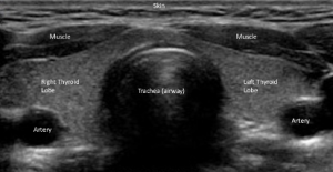
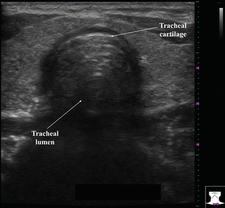
Normal Thyroid Appearance and Anatomic Landmarks in Neck Ultrasound ...
Thyroid ultrasound showing well defined hypoechoic lymph nodes ...
Classic Pattern #8. Hashimoto’s thyroiditis: Longitudinal sonogram ...
| Hydrodissection technique: this technique consists of a pressurized ...
Ultrasound of thyroid gland: (a) thyroid abscess, (b and c) thyroid ...
Thyroid ultrasound disclosed a hypoechoic nodule of 15.5 × 13.5 × 12 mm ...
Image | Radiopaedia.org
Parathyroid adenoma | Radiology Case | Radiopaedia.org Thyroid ...
ECR 2014 / C-0405 / "Mummy what’s this on my neck? - A pictorial review ...
High frequency ultrasonography revealed empty left thyroid fossa ...
Figure 3 from A Child With a Painless Lump on the Anterior Chest Wall ...
Thyroid Cancer Happens | Children's Hospital of Philadelphia
A: USG Right thyroid heterogeneously hypo echoic nodule with ...
Ultrasound of the Thyroid and Soft Tissues of the Neck | SpringerLink
VN (arrow) is located between the IJV and CCA on both sides in ...
Midline neck mass (star) and lateral neck mass (arrow). | Download ...
ULTRASOUND EVALUATION OF RHEUMATOID ARTHRITIS | Musculoskeletal Key
Medullary thyroid carcinoma with nodal metastases | Image | Radiopaedia.org
Ultrasonography longitudinal section of lower neck showing normal right ...
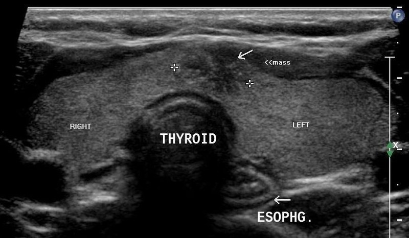
Sagittal image demonstrating a hypoechoic, elongated, complex cystic ...
Chest, Thyroid, Parathyroid, and Neonatal Brain Ultrasound | Radiology Key
Thyroid ultrasound showing well-defined, mildly lobulated, solid with ...
A well-defined, oval shaped, hyperechoic lesion identified in the ...
Interpretation of Ultrasound | Ento Key
Enlarged-Thyroid-Ultrasound
Healthy-Thyroid-Ultrasound
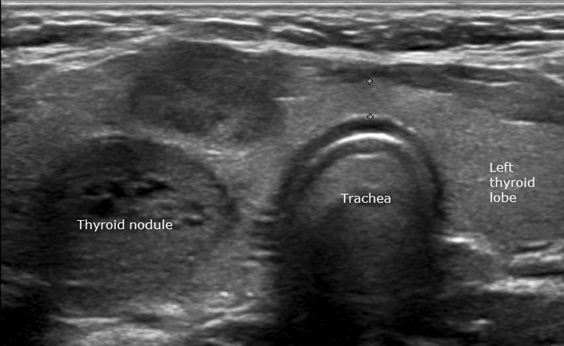
Thyroid-Nodules-Ultrasound
Medullary-Thyroid-Cancer-Ultrasound
Malignant-Thyroid-Nodules-Ultrasound
Neck-Ultrasound-Thyroid
Heterogeneous-Thyroid-Ultrasound
Parathyroid-Ultrasound
Thyroiditis-Ultrasound
Benign-Thyroid-Nodule-Ultrasound
Graves-Thyroid-Ultrasound
Hypoechoic-Thyroid-Nodule-Ultrasound
Us-Thyroid
Thyroid-FNA-Ultrasound
Colloid-Cyst-Ultrasound
Thyroid-Scan