


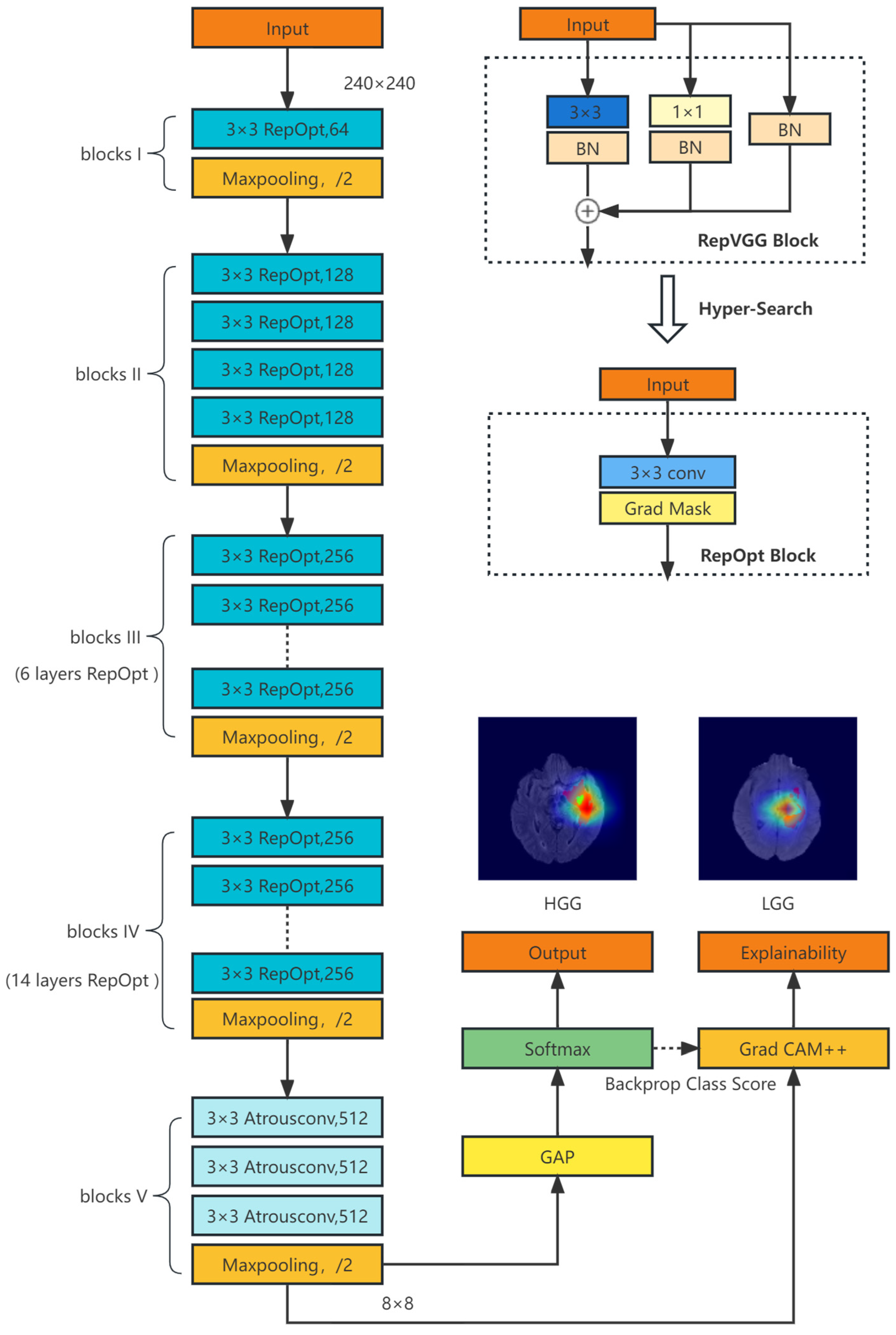


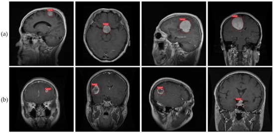


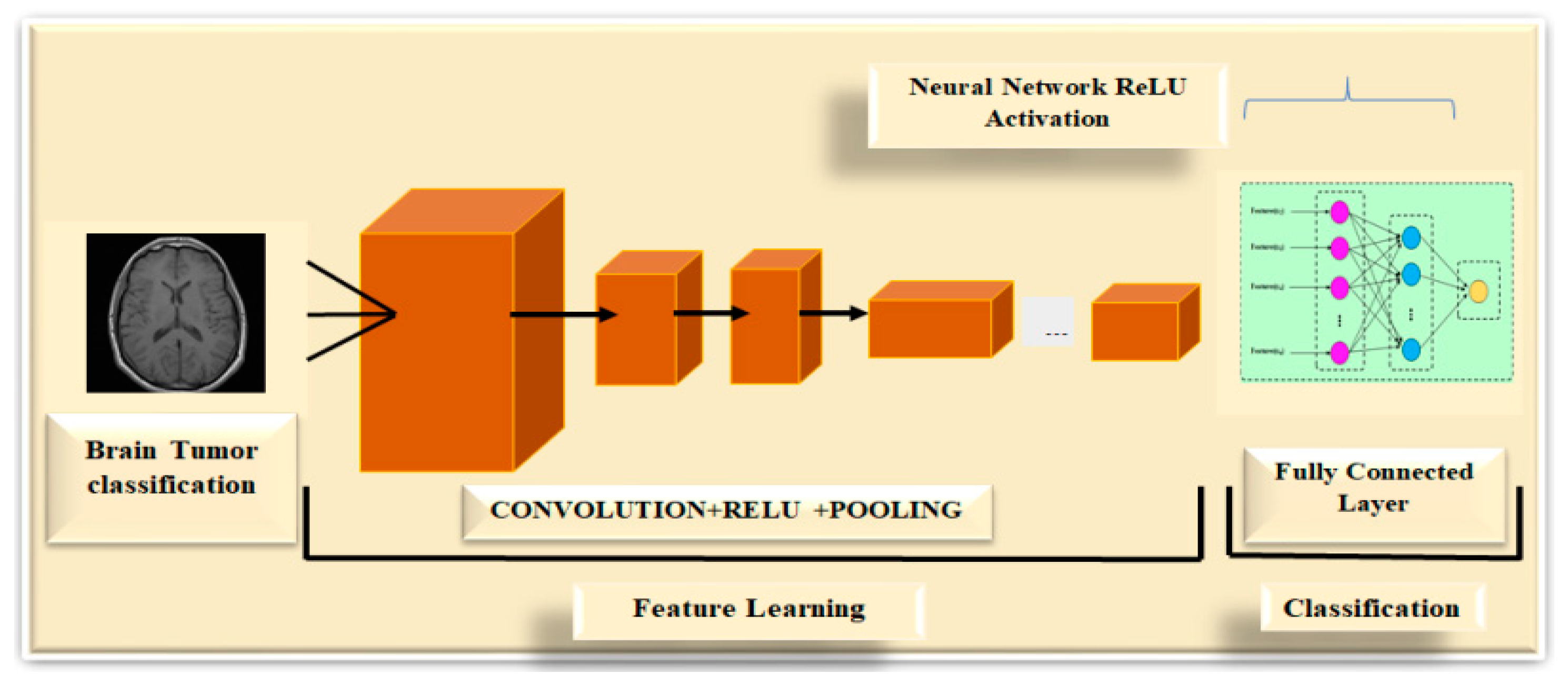


![CNN-based Tumor Classification Another study [79] proposed a BrainMRNet ...](https://www.researchgate.net/publication/374063535/figure/fig2/AS:11431281190344893@1695299017251/CNN-based-Tumor-Classification-Another-study-79-proposed-a-BrainMRNet-as-a-novel.jpg)


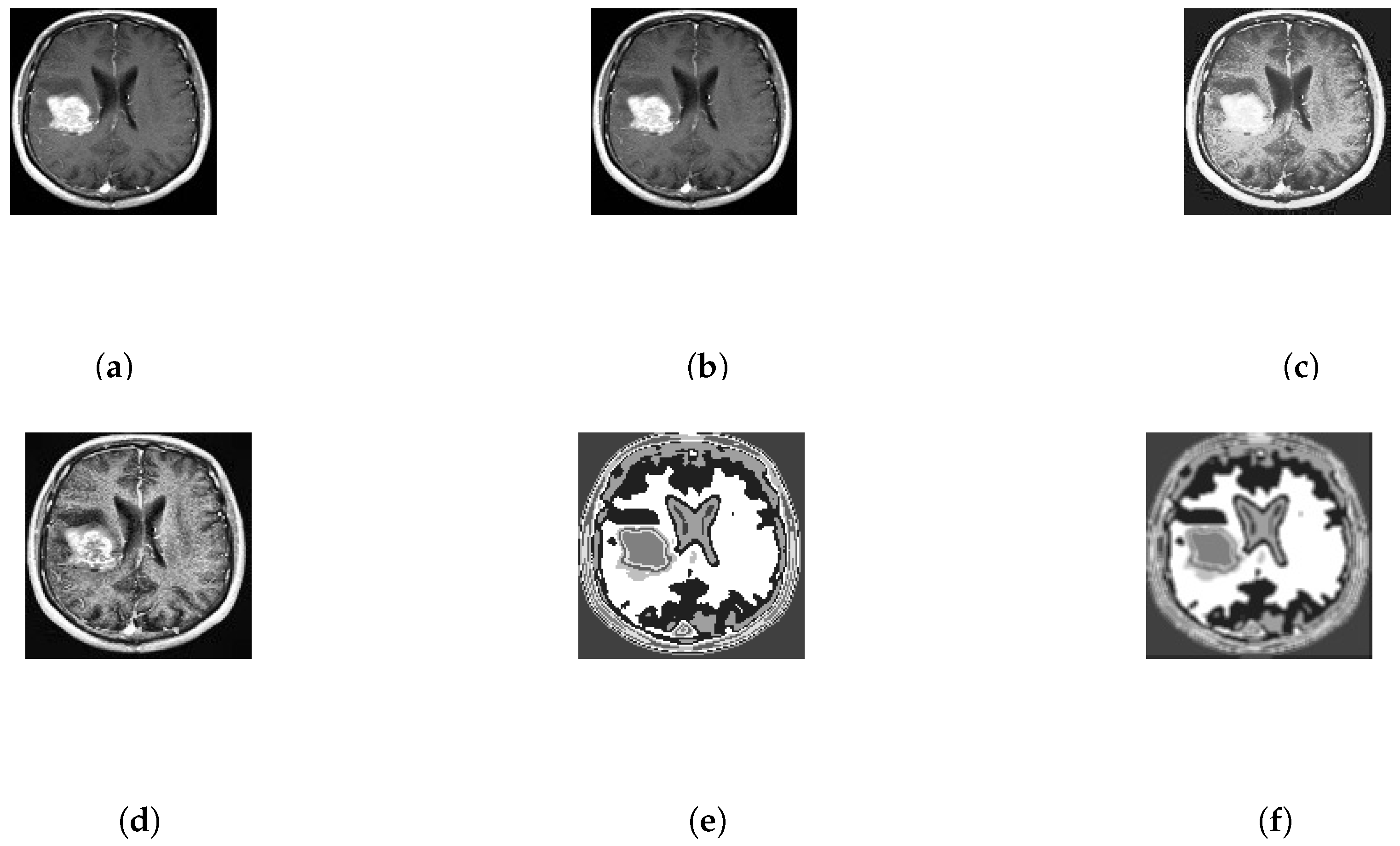


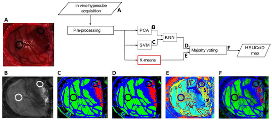


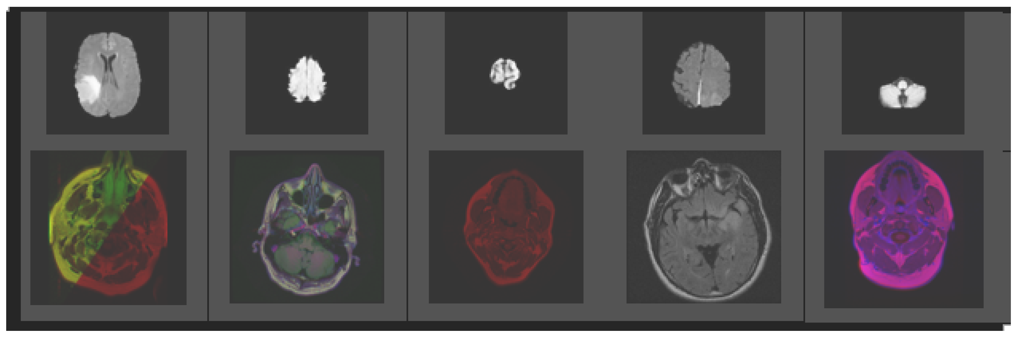
Study the characteristics of figure 1 from brain tumor detection using color-based k-means using our comprehensive set of extensive collections of learning images. providing valuable teaching resources for educators and students alike. encouraging critical thinking and analytical skill development. Discover high-resolution figure 1 from brain tumor detection using color-based k-means images optimized for various applications. Excellent for educational materials, academic research, teaching resources, and learning activities All figure 1 from brain tumor detection using color-based k-means images are available in high resolution with professional-grade quality, optimized for both digital and print applications, and include comprehensive metadata for easy organization and usage. Our figure 1 from brain tumor detection using color-based k-means images support learning objectives across diverse educational environments. Reliable customer support ensures smooth experience throughout the figure 1 from brain tumor detection using color-based k-means selection process. Our figure 1 from brain tumor detection using color-based k-means database continuously expands with fresh, relevant content from skilled photographers. Advanced search capabilities make finding the perfect figure 1 from brain tumor detection using color-based k-means image effortless and efficient. The figure 1 from brain tumor detection using color-based k-means collection represents years of careful curation and professional standards. Whether for commercial projects or personal use, our figure 1 from brain tumor detection using color-based k-means collection delivers consistent excellence.