Please enter url.
Login
Logout
Please enter url.
Lymphomatosis cerebri: a treatable cause of rapidly progressive ...
jnnp.bmj.com
source
Comments
Megalencephaly, polymicrogyria and ribbon-like band heterotopia: A new ...
Brain magnetic resonance imaging (MRI) at presentation (A–C), with ...
MRI findings. a, b MRI findings in patient 1 at the onset of acute ...
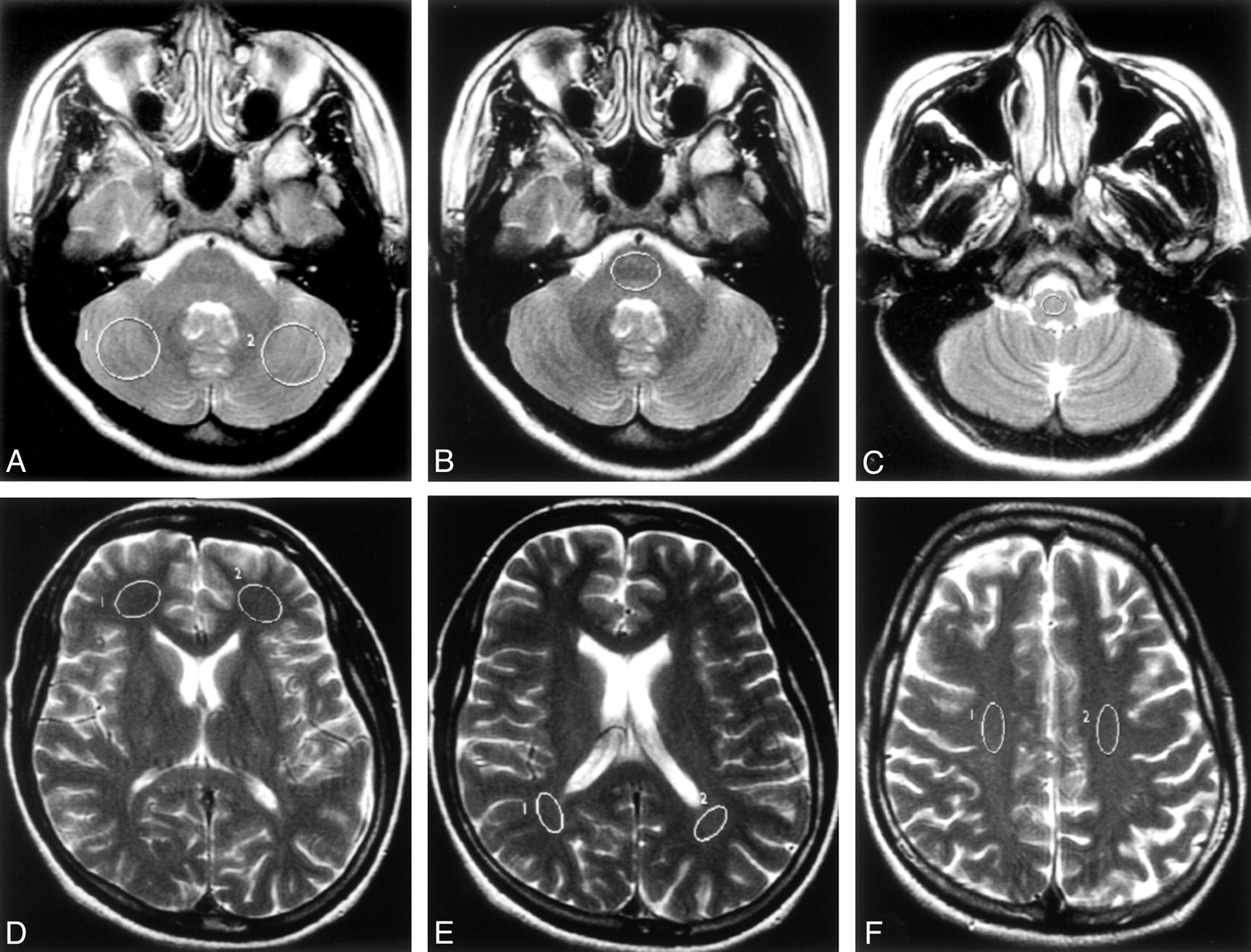
Axial T2 weighted image showing supra- and infra-tentorial high signals ...
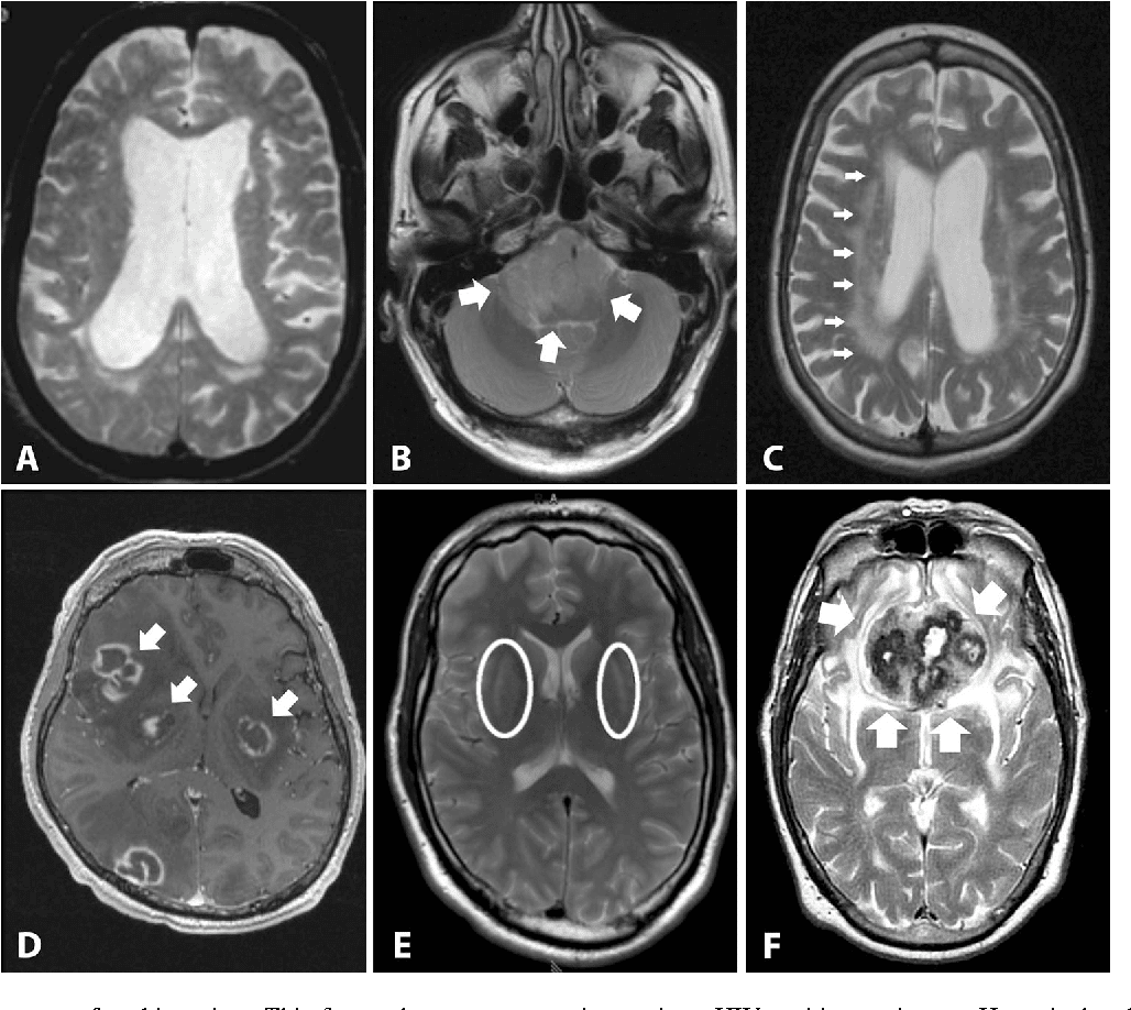
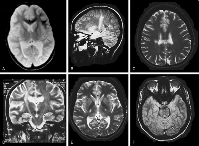
Lymphomatosis cerebri: a rare variant of primary central nervous system ...
Diffuse lesion in the splenium of the corpus callosum in patients with ...
Multimodality Imaging in Primary Progressive Aphasia | American Journal ...
Case 2 at initial presentation. A and B: Axial T2-weighted images show ...
MRI findings of crossed cerebellar diaschisis in a case of Rasmussen’s ...
Baseline brain MRI. (A-C) Multiple patchy foci of diffusion restriction ...
[PDF] Posterior Reversible Encephalopathy Syndrome With Hemorrhagic ...
Preoperative cerebral magnetic resonance imaging (MRI). (a) MRI ...
Parkinson's Disease Mri Findings - ParkinsonsDaily.com
Diffusion-Tensor Imaging for the Detection and Quantification of ...
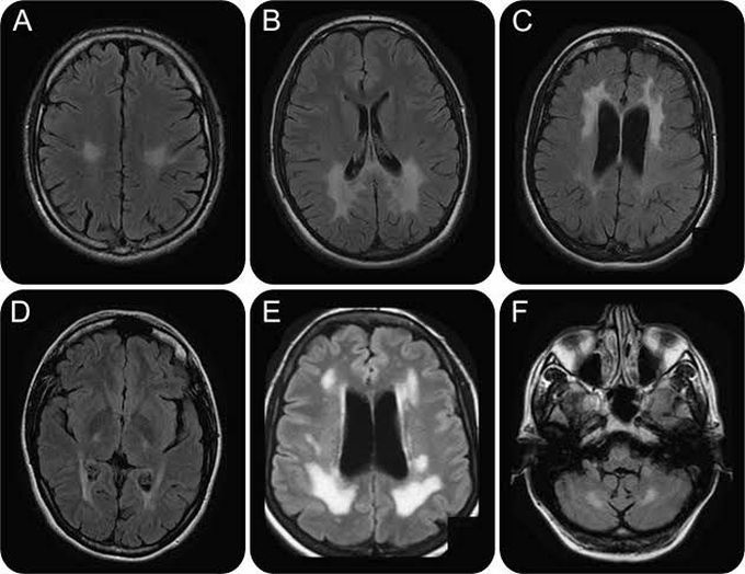
Transient encephalopathy with reversible white matter lesions: A case ...
Neonatal MRI to Predict Neurodevelopmental Outcomes in Preterm Infants ...
A 38-Year-Old Woman With Global Aphasia and Migraine - CHEST
Brain MRI findings in children with Leigh-like syndrome. a Brain MRI ...
MRI showing iron in BPAN. The globus pallidus is variably hypointense ...
PINK, PANK, or PARK? A clinicians' guide to familial parkinsonism - The ...
Patient 1. Axial non-contrast-enhanced CT brain scan revealing a left ...
Brain MRI imaging. T2-weighted TSE axial scans (1.5 T) at the level of ...
Krabbe Disease - MEDizzy
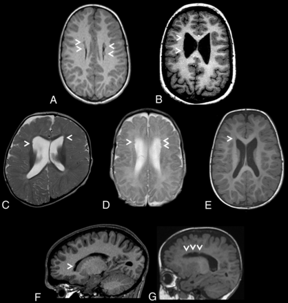
Distinctive Brain Malformations in Zhu-Tokita-Takenouchi-Kim Syndrome ...
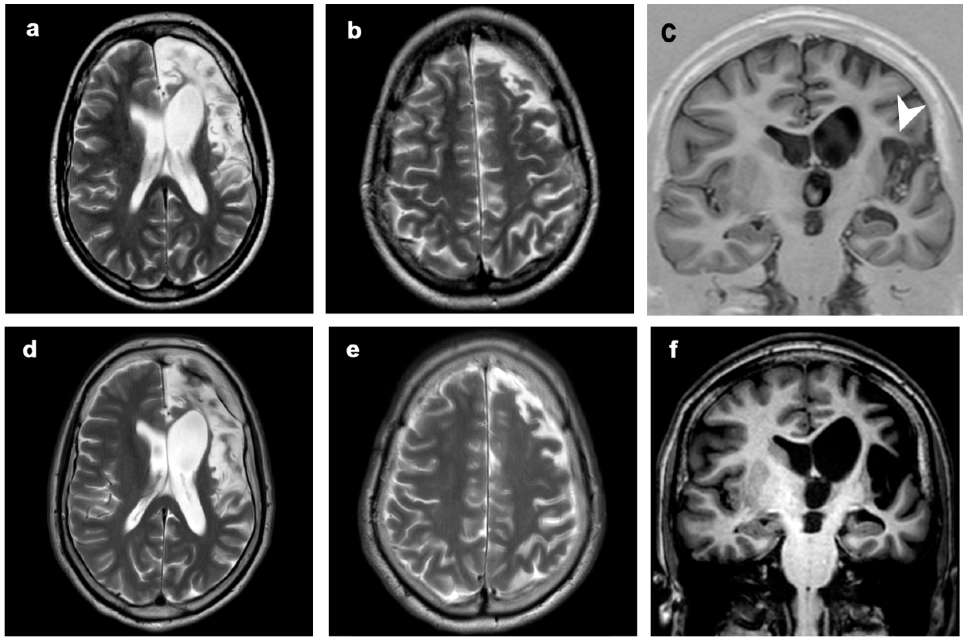
Representative MRI findings in different malformations of cortical ...
MRI in CLN2 disease patients: Subtle features that support an early ...
T2-weighted MRI images of patient 7, taken at the age of 19 months ...
Bilateral basal ganglia lesions at presentation (a–e): hypodense on CT ...
THE LEUKODYSTROPHIES | Neupsy Key
Bilateral occipital cortical dysplasia and white matter T2 ...
Diagnostics | Free Full-Text | MRI in Late-Onset Rasmussen Encephalitis ...
Figur 1 Pasient med terapiresistent epilepsi som har vaert undersøkt ...
(PDF) mTOR Pathway Mutations Cause Hemimegalencephaly and Focal ...
Cerebral Amyloid Angiopathy: Diagnosis, Clinical Implications, and ...
Hypoglycemic encephalopathy mimicking acute ischemic stroke in clinical ...


![[PDF] Posterior Reversible Encephalopathy Syndrome With Hemorrhagic ...](https://d3i71xaburhd42.cloudfront.net/42553b791acc17880d138fae3a36691f1cd77af4/2-Figure1-1.png)