Please enter url.
Login
Logout
Please enter url.
What Is Transitional Cell Carcinoma In Dogs
animalia-life.club
source
Comments
Canine transitional cell carcinoma of the urinary bladder: a focus on ...
fig2:Rapidly Growing Thyroid Mass in an Immunocompromised Young Male ...
Thyroid ultrasound: (a) left midlobe dominant nodule in transverse view ...
Implant-associated anaplastic large cell lymphoma | Eurorad
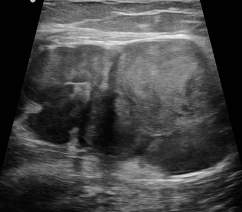
Pediatrics | 9.1 Pediatric abdomen and retroperitoneum : Case 9.1.10 ...
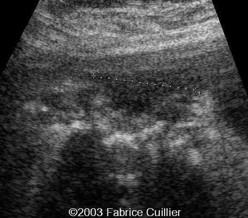
Uretropelvic junction obstruction in utero | Eurorad
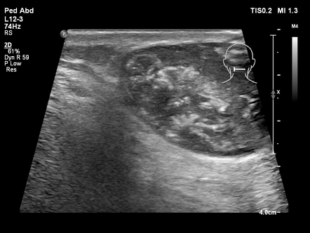
b. Well bordered complex solid-cystic mass lesion from a different ...
(PDF) Can you spot diverticulitis in the ultrasound?
A Gallery of High-Resolution, Ultrasound, Color Doppler & 3D Images ...
Pilomatricoma | Radiology Case | Radiopaedia.org
Tissue air bubbles in necrotising fasciitis without gas producing ...
Ultrasound neck showing normal thyroid gland and no abnormal ...
[PDF] Sonoelastography Findings of Breast Juvenile Papillomatosis: A ...
Cardiac manifestations of anaphylaxis in dogs – Vet Practice Support
Left male breast cancer presenting with breast lump (scar of biopsy is ...
Images | Radiopaedia.org
16 year old male with high blood pressure for over 2 years diagnosed ...
Ecografía que muestra la masa. | Download Scientific Diagram
EPOS™
Patient 2. (a) Photograph of the physical features of the patient with ...
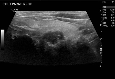
Hyperparathyroidism | Radiology Reference Article | Radiopaedia.org
Renal infarcts in dogs: a few images – Vet Practice Support
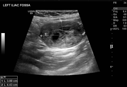
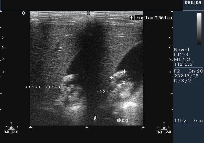
Abdominal ultrasound: The appendix is enlarged (diameter 1 cm) and ...
Radiology case: Metastatic infiltration of lymph nodes
Aneurysm of the abdominal aorta ultrasound longitudinal … | Ultrasound ...
A case of testicular filariasis, causing echogenicity quite different ...
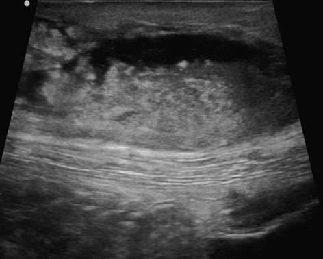
Ultrasound imaging: large left sided mass with inhomogeneous tissue ...
📃 Spina bifida aperta
A surgical soft tissue surprise | Eurorad
The left solid cyst of the thyroid gland was injected with 10mL of ...
Junctional parenchymal defect of kidney | Radiology Reference Article ...
Lymph Nodes Ultrasound | pictureperfectus
Musculoskeletal, bone, muscle, nerves and other soft tissues | 7.5 Soft ...
Right testicular ultrasound showing a 4.0 cm section of the 5.5×3.5×2.5 ...
Figure 2 from Ultrasound Study to Validate the Anterior Cervical ...


![[PDF] Sonoelastography Findings of Breast Juvenile Papillomatosis: A ...](https://d3i71xaburhd42.cloudfront.net/28a8b5f8b7b537aea2acc82a6419d4ef79bf681c/2-Figure1-1.png)