Please enter url.
Login
Logout
Please enter url.
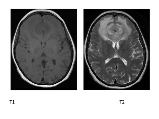
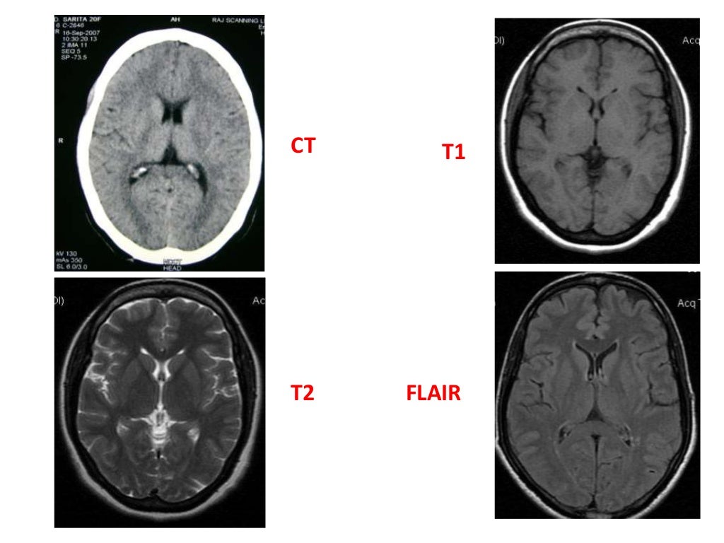
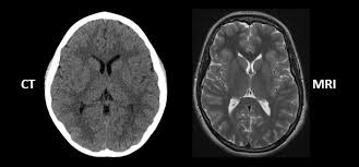
Mri Vs Ct Brain
mavink.com
source
Comments
Durable remission with Bruton’s tyrosine kinase inhibitor therapy in a ...
Diagnostic Imaging of Brain Tumors | PPT
Routine clinical brain MRI sequences for use at 3.0 Tesla - Lu - 2005 ...
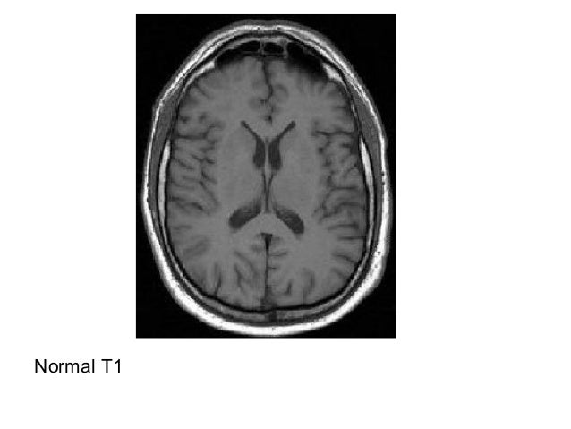
Normal mri brain
Effect Of Magnetic Resonance Imaging - Non-Ionizing Radiation in MRI
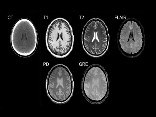
MRI Sequences in Neuroradiology
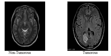
Electronics | Free Full-Text | A Novel Approach for Classifying Brain ...
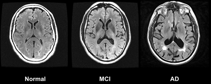
Magnetic resonance imaging in Alzheimer’s disease and mild cognitive ...
MRI CLINICAL APPLICATIONS
MRI of a 22-year-old woman affected by Streptococcus pneumoniae ...
72-year-old male with a history of squamous cell carcinoma of the base ...
Journal-Neurological-Disorders-Brain-MRI
LMC in contrast-enhanced MRI.a Cranial nerve enhancement. b Folia ...
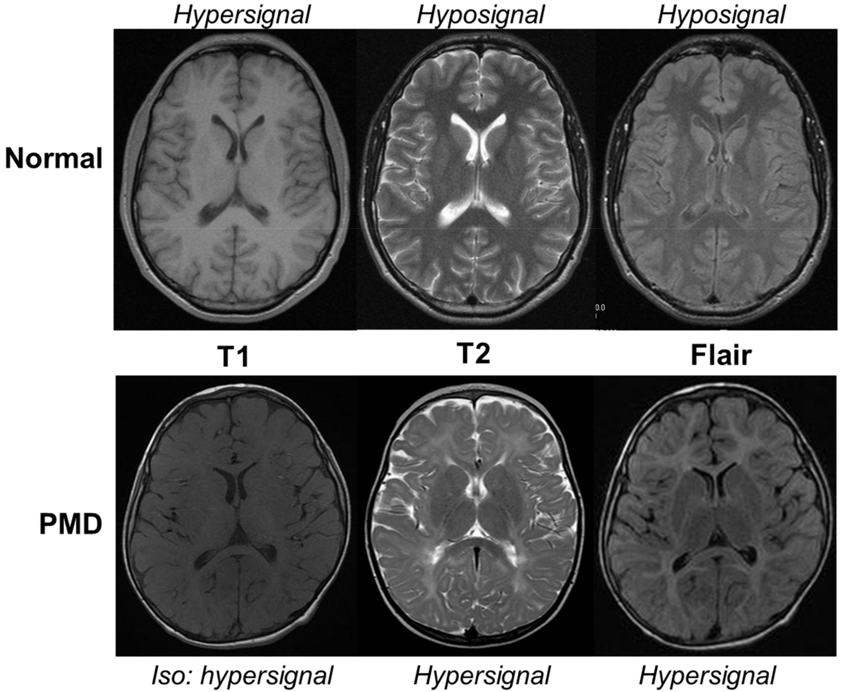
MRI basics - How to read and understand MRI sequences
Ischemic: Frontal Lobe Ischemic Stroke
Brain Tumor Detection
Brain MRI scans showed slight changes: choroid plexus cysts in the ...
Uncommon and atypical meningiomas and imaging variants: A report of 7 cases
Meningoencephalitis or meningitis in relapsing polychondritis: Four ...
Brain MRI (T1-and T2-weighted, axial images). (a, b) Patient 1 at age 1 ...
Linear scleroderma “en coup de sabre” with extensive brain involvement ...
Oncotarget | Perioperative cerebral ischemia promote infiltrative ...
SciELO - Brasil - GM1 gangliosidosis: a case report GM1 gangliosidosis ...
Cortical gray matter lesions in acute encephalopathy with febrile ...
Axial PD-weighted ( A ), T 2 -weighted ( B ) and T 1 -weighted ( C ...
Frontiers | Assessing Performance on Digital Clock Drawing Test in Aged ...
Imaging in Fahr’s disease: how CT and MRI differ? | BMJ Case Reports
Clinical variability and the role of diagnostic criteria of cerebral ...
Frontiers | Superficial Siderosis and Microbleed Restricted in Cortex ...
Reanalysis of exome sequencing data reveals a treatable neurometabolic ...
Two patients with cerebral lesions: is it tumor or multiple sclerosis ...
Biomedicines | Free Full-Text | Mutation of Proteolipid Protein 1 Gene ...
Follow up MR image of brain done after 4 weeks. FLAIR axial Aand T2 ...
Imaging in Fahr’s disease: how CT and MRI differ? | BMJ Case Reports
Pin on Lipoma Health
MRI-X-ray
Cat-Scan-Stroke
CT-vs-X-ray
MRI-vs-CT-Scan-Machines
X-ray-Brain-Scan
Brain-Imaging
Mini-Stroke-CT-Brain-Scan
Cat-Scan-Brain-Tumor
CT-Scan-Head-Stroke
CT-Scan-Images-of-Brain
Brain-CT-Scanner
CT-Scan-Radiation
Cat-Scan-versus-MRI
Pet-MRI-Scan
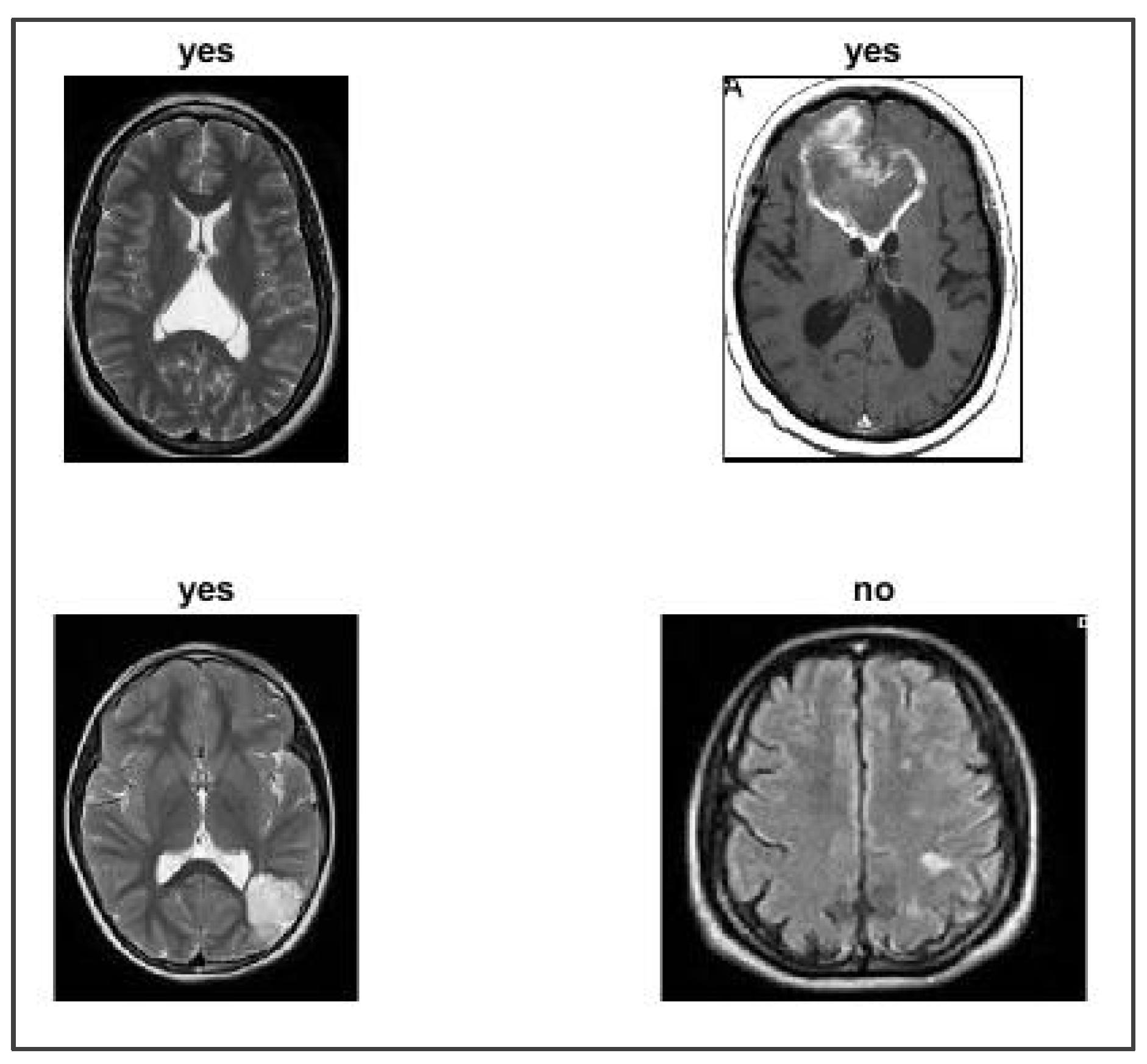
Abnormal-Brain-MRI
Brain-CT-Scan-with-Contrast