Please enter url.
Login
Logout
Please enter url.
Kidney Ultrasound Tumor
ar.inspiredpencil.com
source
Comments
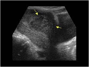
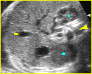
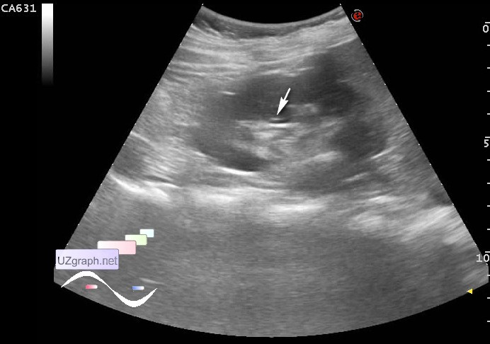
Normal right kidney in longitudinal view. Arrow: cortex, star ...
Abdomen and retroperitoneum | 1.9 Retroperitoneum and great vessels ...
Microbubble-enhanced US in Body Imaging: What Role? | Radiology
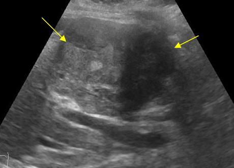
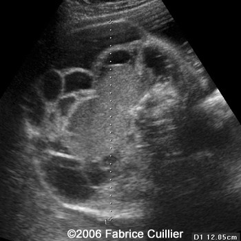
Ultrasound imaging: Sonography of a case of bilateral fetal hydronephrosis
Abdomen and retroperitoneum | 1.1 Liver : Case 1.1.7 Hepatocellular ...
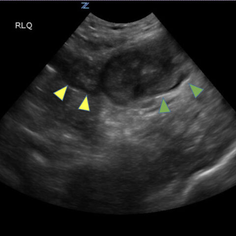
Sonoguide // Renal Ultrasound
Hepatosplenomegaly – Department of Obstetrics and Gynecology Faculty of ...
b: A 21 year old female patient with uterine perforation and abdominal ...
Abdomen and retroperitoneum | 1.1 Liver : Case 1.1.7 Hepatocellular ...
Lethal multiple pterygium syndrome: A severe phenotype associated with ...
Pediatric Ovarian Torsion: Spectrum of Imaging Findings | RadioGraphics
Imaging Findings of Small Bowel - Diverticulitis: A Case Report - JETem
Image | Radiopaedia.org
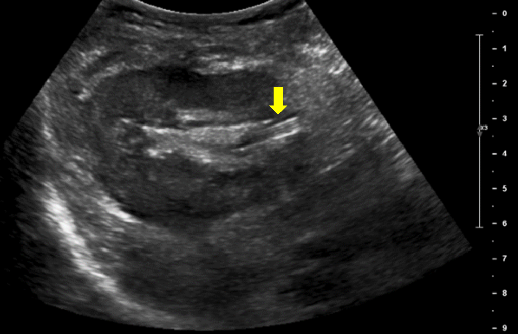
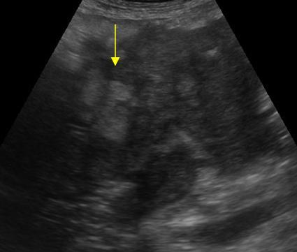
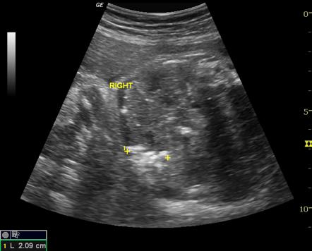
Longitudinal view of right kidney with hyperechoic structure in the ...
GB agenesis, Choledochal cysts, urolithiasis - Abdomen sonography ...
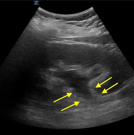
Pre-treatment ultrasound of graft kidney with ureter showing mild ...
Ultrasound imaging: Sonography of a case of bilateral fetal hydronephrosis
POCUS Gallery - Renal Fellow Network
Imaging Findings of Small Bowel - Diverticulitis: A Case Report - JETem
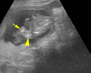
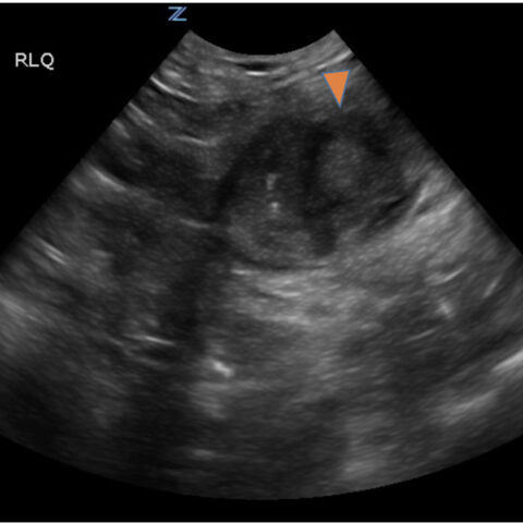
Abdominal ultrasound showing a 6.2 cm cyst at the upper pole of the ...
Posterior urethral valves: not uncommon presentation in older age ...
Gynaecology | 3.1 Uterus : Case 3.1.3 Malignant uterine and cervical ...
Limb Reduction Defects – Department of Obstetrics and Gynecology ...
Point-of-care ultrasonography of the right upper quadrant, identifying ...
Elevated right hemidiaphragm | Eurorad
(PDF) Diagnosis of acute pyelonephritis by contrast-enhanced ...
Image | Radiopaedia.org
Sagittal right upper quadrant sonogram shows numerous hypoechoic ...
Abdomen and retroperitoneum | 1.1 Liver : Case 1.1.7 Hepatocellular ...
1000+ images about Urology x-ray on Pinterest | Stones and Columns
📃 Cervical lymphangioma
Nuchal Thickening – Department of Obstetrics and Gynecology Faculty of ...
| Grade I GMH-IVH. (A-C) Ultrasound scan in a preterm infant (GA 26 ...
US of Gastrointestinal Tract Disease | RadioGraphics
Now You See It, Now You Don’t: Visual Illusions in Radiology ...
Inside-a-Kidney
A-Diagram-of-a-Kidney
Kidney-Profile
Picture-of-Real-Kidney
Normal-Kidney-CT
Healthy-Kidney-Picture
Kidney-Area
Chronic-Kidney-Stages
Kidney-Model
Kidney-Function
Normal-Kidney-Size-Chart
Kidney-Illustration
Signs-of-Kidney-Disease
Normal-Kidney-Cross-Section
Basic-Kidneys
Kidney-Model-Labeled


.jpg)


.jpg)