Please enter url.
Login
Logout
Please enter url.
Cureus | Diagnostic Yield of CT Pulmonary Angiogram in the Diagnosis of ...
cureus.com
source
Comments
Cureus | Diagnostic Yield of CT Pulmonary Angiogram in the Diagnosis of ...
Osteolytic metastatic lesions of the left fourth rib (A) and the fourth ...
Repeat enhanced thoracic CT prior to initiation of cladribine therapy ...
Group A Streptococcus necrotising myositis of the limbs secondary to ...
Balloon pulmonary angioplasty – efficient therapy of chronic ...
(A-G) Computed tomography (CT) of a 60-year-old male on prolonged ...
Computed tomography (CT) scan of the lungs before and after treatment ...
Map showing the endemic areas for histoplasmosis and blastomycosis in ...
Incidentally Found Aorto-Pulmonary Middle Mediastinal Hypervascular ...
The image changes before and after treatment. The CT scan before ...
A female patient 47 years old presented with dyspnea and chest pain ...
Massive Pulmonary Thrombosis Following Haemoptysis in Type IV Ehlers ...
Figure 1 from Diffuse Pleural Myeloid Sarcoma Mimicking Malignant ...
Response to First-Line Osimertinib Treatment in Non–Small-Cell Lung ...
Association between pulmonary embolism and COVID-19 severe pneumonia ...
-Contrast cardiac computed tomography image of the lung window (right ...
(A) February 3, 2020 CT Thorax: Right paratracheal adenopathy, axial ...
Examples of CTPA of COVID-19 patients with acute pulmonary embolism ...
Imaging Features of Primary Immunodeficiency Disorders | Radiology ...
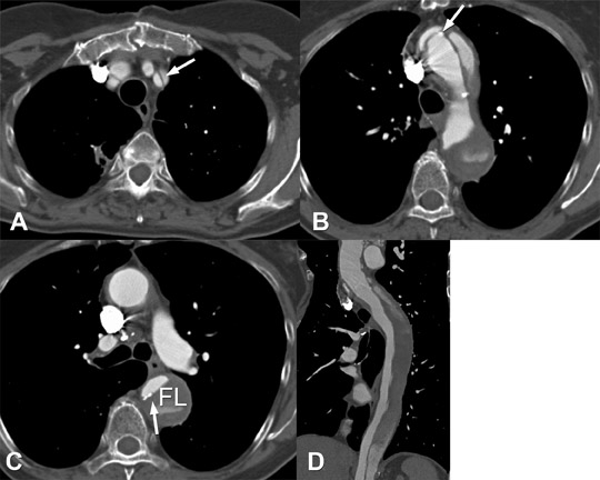
Thoracic aorta: Acute Syndromes • APPLIED RADIOLOGY
(A-D) 2014-9-9 chest CT showed patchy infiltration in the left lower ...
Thoracic CT with contrast revealed Fig. 1A. Narrowing of proximal ...
Imaging of the bronchial arteries. CECT findings of normal bronchial ...
Representative microphotographs showing hypercortisolemia-related ...
White grass line illustrates the quantitative measurements performed on ...
Frontiers | Long-term, 13-year survival after immune cell therapy ...
Chest CT angiogram during the second hospitalization A: right pulmonary ...
Long-term progression-free survival of third-line apatinib in lung squ ...
An Integrated Radiologic-Pathologic Understanding of COVID-19 Pneumonia ...
A 57-year-old man with moderate COVID-19 pneumonia. (A, B) The first ...
Cardiac MRI Anatomy MRIquiz Flashcards | Quizlet
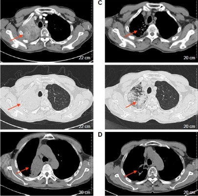
Tumors invading SVC on chest CT. a Right hilus pulmonis Sq.in patient ...
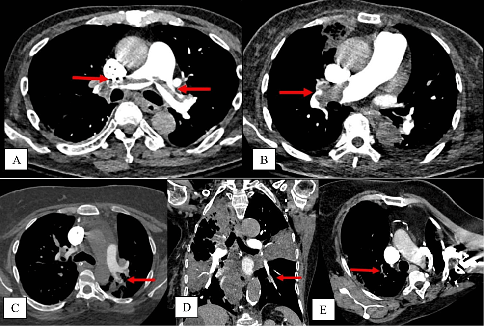
CT pulmonary angiography showing multiple emboli in the main & distal ...
Lymphadenopathy in a 15-year-old boy with human immunodeficiency virus ...
Persistent left superior vena cava in CT in 3D-reconstruction. A ...
Pulmonary-Angiogram-Anatomy
Pulmonary-Artery-Angiogram
Pulmonary-Embolism-Angiogram
Pulmonary-Angio
CT-Angio-Pulmonary-Embolism
VQ-Scan-Pulmonary-Embolism
Lung-Pulmonary-Embolism
Pulmonary-Embolism-ABG
Extensive-Pulmonary-Embolism
Pe-Angiogram
Distal-Pulmonary-Embolism
AngioJet-for-Pulmonary-Embolism
Segmental-Pulmonary-Embolism
CT-Angio-Bilateral-Pulmonary-Embolism
Acute-Pulmonary-Embolism
Pulmonary-Arteries-Segments