Please enter url.
Login
Logout
Please enter url.
Low-frequency stimulation of a fiber tract in bilateral temporal lobe ...
epilepsybehavior.com
source
Comments
Hemi-seesaw Nystagmus in Joubert Syndrome | Canadian Journal of ...
(A). Patient interictal electroencephalogram (EEG) and... | Download ...
Unilobar surgery for symptomatic epileptic spasms - Barba - 2017 ...
Clinical and IONM results. a MR imaging. Frontal and axial images...
(Case 2) (a) A CT scan revealed multiple contusional and subarachnoid ...
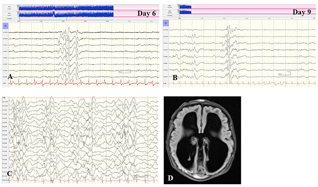
Frontiers | Neuromonitoring in Neonatal-Onset Epileptic Encephalopathies
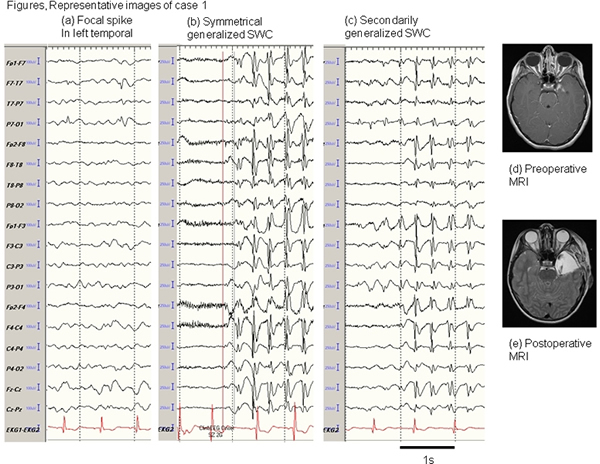
Generalized-3-Hz-spike-and-wave-complexes-emanating-from-focal ...
Examples of interictal spikes in Case 1. ( Left ) Interictal spike ...
Detection of glutamate, glutamine, and glutathione by radiofrequency ...
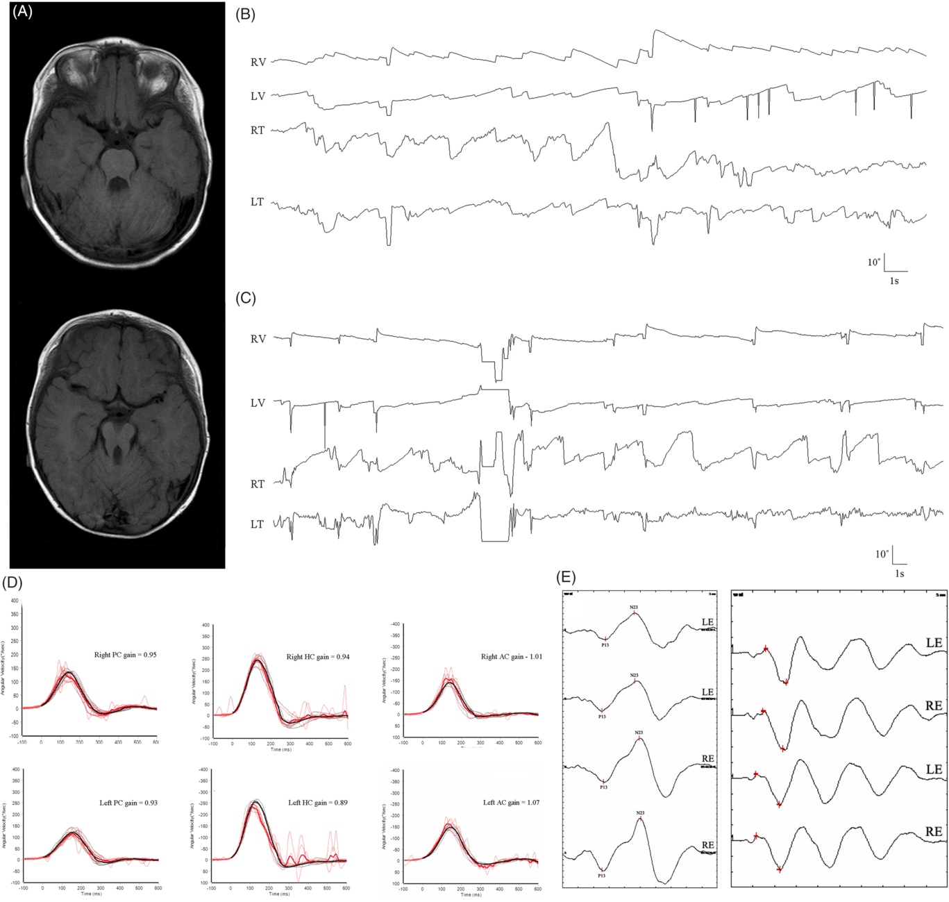
Figure 1 from A Unique Shape of Brainstem Lesion that Caused ...
(a) A telemetry electroencephalograpy (EEG) amplifier (AE-120A EEG ...
Figure 1 from Median nerve SEP after a high medullary lesion. Preserved ...
Postictal psychosis: Evidence for extrafocal functional precursors ...
Isolated dysarthria due to extracerebellar lacunar stroke: a central ...
Focal magnetoencephalographic spikes in the superior temporal plane ...
A: MRI showed a left frontal (premotor) lesion reminiscent to a tumor ...
De novo mutation in DEPDC5 associated with unilateral pachygyria and ...
Electroencephalogram (EEG) and magnetic resonance imaging (MRI) in ...
Occipital lobe seizures related to marked elevation of hemoglobin A1C ...
Artifacts | Neupsy Key
MRI images (upper right) show a large arachnoid cyst in the left ...
Periodic lateralized epileptiform discharges (PLEDs) in herpetic ...
The dysplastic areas of Case 1. (A) The left temporal dysplasia ...
A girl with early-onset epileptic encephalopathy associated with ...
Typical spectra from the tumor and control groups. Typical 1 H-MRS and ...
[A]: An intraoperative photograph shows the cloudy arachnoid membrane ...
Epilepsy Surgery Outcome in Coexisting Symptomatic Refractory Focal ...
Images from the same case of MELAS-MERRF overlapping syndrome shown ...
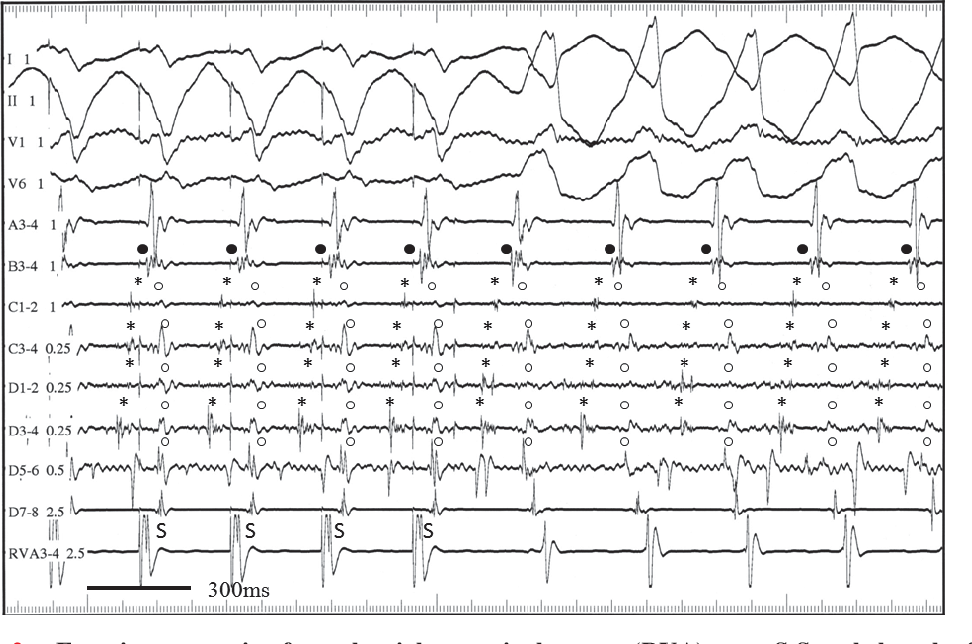
Figure 2 from Onset of reentrant ventricular tachycardia modulated by ...
(Case 4) (a) A CT scan revealed an old contusion in the left frontal ...
A case of drug-resistant epilepsy and autism with de novo SLC6A8 gene ...
Examples of SEEG signals recorded in different tissues. Pre-(only a and ...
Neurophysiological exam of the boy at 3 years of age, performed during ...
A: MRI showed a left frontal (premotor) lesion reminiscent to a tumor ...


![[A]: An intraoperative photograph shows the cloudy arachnoid membrane ...](https://www.researchgate.net/publication/236655151/figure/fig2/AS:299287960408071@1448367218763/A-An-intraoperative-photograph-shows-the-cloudy-arachnoid-membrane-on-the-Sylvian.png)