Please enter url.
Login
Logout
Please enter url.
source
Comments
CF8
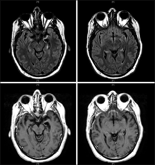
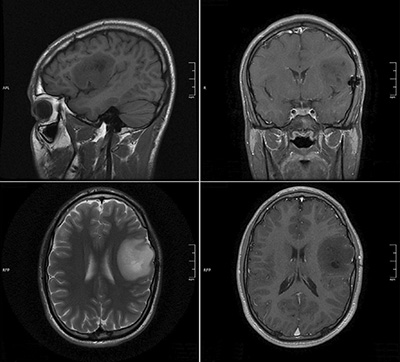
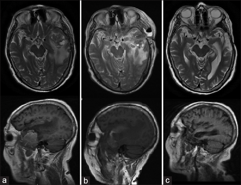
MRI of the brain with contrast showing minimal scattered nonspecific ...
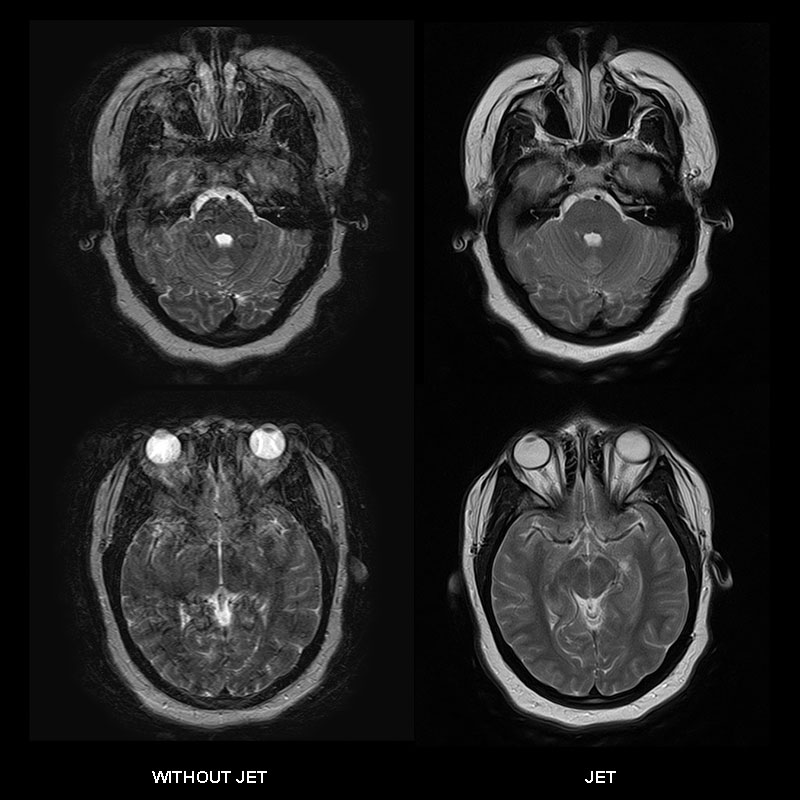
Head and Neck Clinical Gallery | Vantage Elan 1.5T | Magnetic Resonance ...
Neurologic Complications After Kidney Transplantation - Seminars in ...
Brain Meningioma Imaging: Practice Essentials, Radiography, Computed ...
Dentate Nucleus T1 Hyperintensity in Multiple Sclerosis | American ...
Frontiers | Case Report: Detection of SARS-CoV-2 From Cerebrospinal ...
Figure 1 from HIV positive patient with HSV-2 encephalitis: case report ...
Control magnetic resonance imaging after surgery, chemotherapy and ...
MRI FLAIR sequence. Part 1 and 2 Pretreatment images shows hyperintense ...
Archives Skull Base Meningioma Embolization MHT Access | neuroangio.org
Crossed cerebellar diaschisis: a radiological finding in status ...
Figure 4:A Case of Cerebral Erdheim-Chester Disease With Progressive ...
Surgical Neurology International
Figure MRI of demyelinating lesions after etanercept therapy | Download ...
Brain Cancer Expert Joins Allegheny Health Network | Highmark Health Blog
Cerebral Infarction from Compression of the Internal Carotid Artery - A ...
(PDF) Management of pseudomeningocele following posterior fossa tumor ...
Case 1. T 1 -weighted magnetic resonance images with contrast medium ...
August 2021 - Case of the Month | American Journal of Neuroradiology
MRI scans for the quadruped man. Left: sagittal sections, above normal ...
(PDF) Large epidermoid cyst of the cavernous sinus: Case report
Clippers Syndrome - Case 212 Chronic Lymphocytic Inflammation With ...
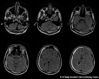
Dr Balaji Anvekar FRCR: Crossed cerebellar diaschisis MRI
MRI manifestations of hypotension syndrome - iMedia
Measurement of a suprasellar component of pituitary adenoma. a, line ...
Preoperative magnetic resonance images show the fourth ventricular ...
Preliminary experience in the management of brain and skull-base tumors ...
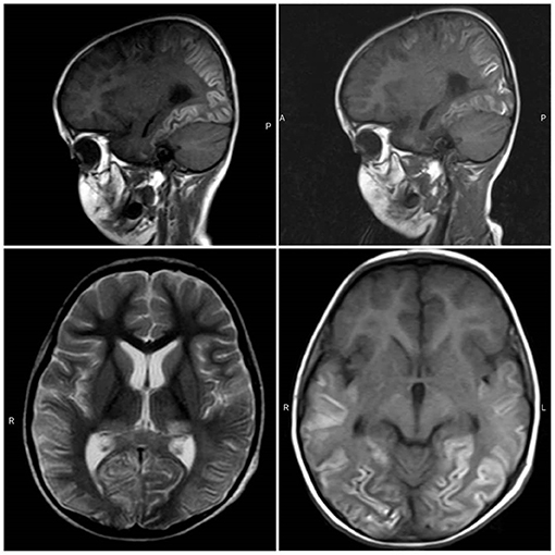
(PDF) Epilepsia estructural por síndrome de Dyke Davidoff Masson: un ...
Surgical Neurology International
T2-weighted MRI of the brain. The MRI on the left is from 2005, and the ...
A: Initial brain MRI showed hyperintensity with slight edema on the ...
Postoperative MRI after 3 months. (Top left) Sagittal T1 image shows no ...
Anti-GAD antibodies, a rare cause of limbic encephalitis: a case report ...
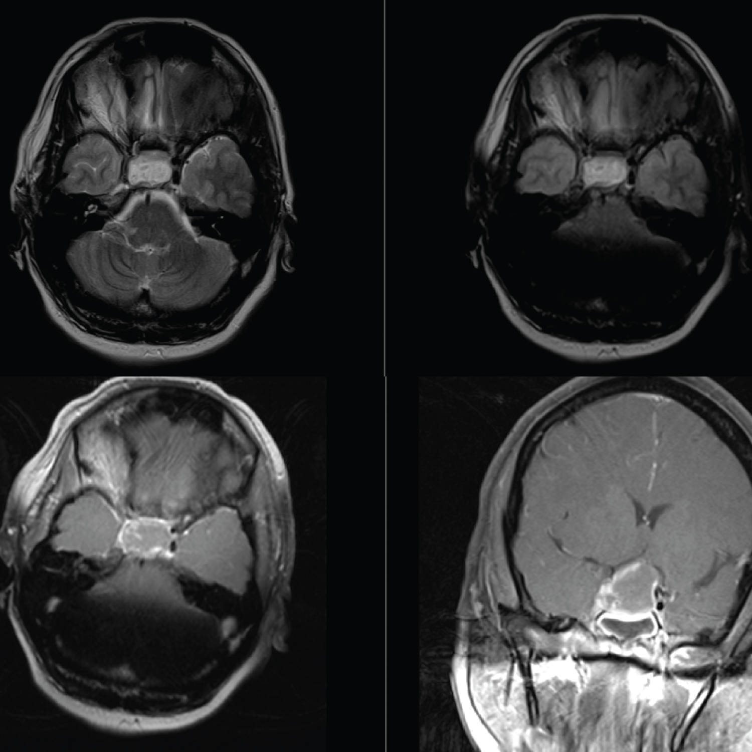
Neuroradiology On the Net: Cerebellar pontine angle meningioma
Central pontine myelinolysis in a patient with non‐Hodgkin lymphoma ...
Multiple-Sclerosis-Brain-MRI-with-Contrast
MRI-Brain-Scan-with-Contrast
Stroke-Brain-MRI-with-Contrast
MS-Brain-MRI-with-Contrast
Brain-Tumor-MRI-Contrast
Brain-MRI-Procedure
MRI-Brain-Imaging
MRI-Brain-Scan-without-Contrast
Sagittal-MRI-Brain
MRI-Brain-Pituitary
MRI-Brain-Anatomy
MRA-Brain-Aneurysm
Abnormal-Brain-MRI-without-Contrast
T1-MRI-Brain
White-Lesions-On-Brain-MRI
Normal-and-Abnormal-Brain-MRI