Please enter url.
Login
Logout
Please enter url.
Patient and Physician Perspectives in a Case of Severe Traumatic Brain ...
link.springer.com
source
Comments
Patient and Physician Perspectives in a Case of Severe Traumatic Brain ...
Ischemic infarct detection, localization, and segmentation in ...
Axial CT scan without injection brain images. Cranial CT scan imaging ...
Suggested algorithm for managing acute liver failure and hepatic ...
CT of the brain without contrast within normal limits (a-e) Views of ...
ECG on admission showing diffuse ST segment depressions ECG ...
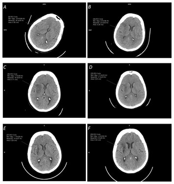
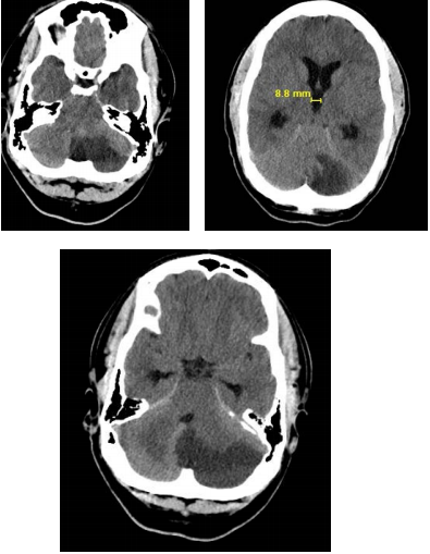
CT scan brain trauma survey for traumatic brain injury in LMS case. a ...
Computerized tomography scans of representative cases with chronic ...
Neuroimaging Classification of Traumatic Brain Injury | Radiology Key
Post-traumatic cerebral venous sinus air embolism | Journal of ...
Frontiers | Romiplostim for the Emergency Management of Severe Immune ...
(a) CT image and (b) MRI image. | Download Scientific Diagram
Hydrocephalus in Children and Adults | Neupsy Key
CT scans of PB patients in the acute stage. Although lesion data do not ...
Anoxic brain injury CT and MRI patterns - quick pictoral quide for ...
CT imaging of the head CT of the head showed no evidence of hemorrhage ...
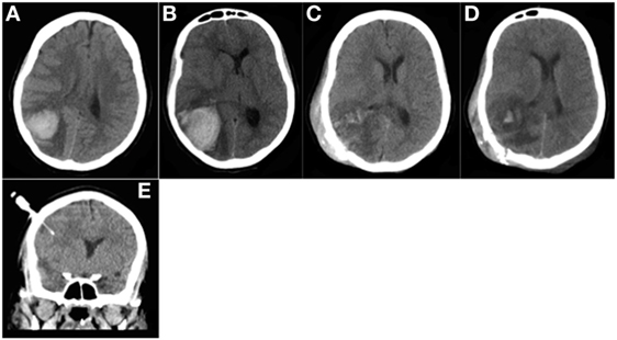
CT scan of the head (a: transverse, b: coronal, c: sagittal) showing ...
Neoplastic And Infectious Aneurysms | MedLink Neurology
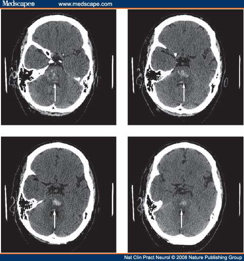
Posterior Inferior Cerebellar Infarct in a Younger Adult Male with ...
Figure 1 from Neuroendoscopic Removal of Acute Subdural Hematoma with ...
Figure 1 from HEMATOMA SUBDURAL CRÓNICO EN PACIENTE CON QUISTE ...
Case report 2. a , b Preoperative CT scan depicting slit-like ...
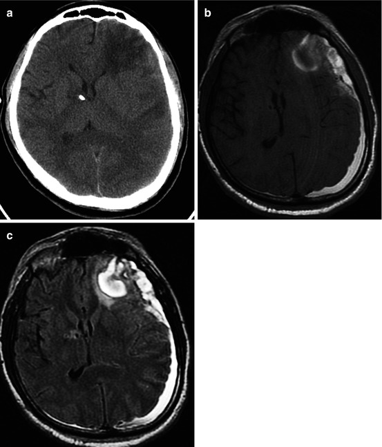
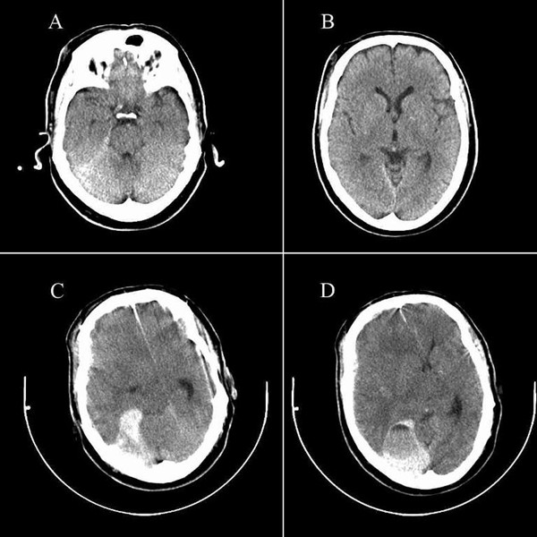
(A) Brain computed tomography (CT) scan at the time of admission ...
Brain | SpringerLink
Patterns of cerebral infarction on computed tomography scans: (A ...
(PDF) Repeated Dosing of 23.4% Hypertonic Saline for Refractory ...
Delayed life-threatening subdural hematoma after minor head injury in a ...
(a) Preoperative computed tomography (CT) of the brain showing ...
Figure 1 from Title Intracranial Myeloid Sarcoma Metastasis Mimicking ...
Preoperative head computed tomography (CT) scan. This axial ...
Representative CT images of the collateral circulation grading. a Good ...
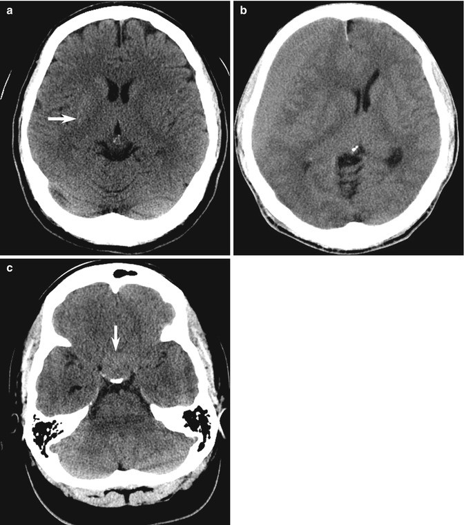
3 days of follow-up brain CT scans showing the progression of bilateral ...
-Ischemic (panel A) and hemorrhagic (panel B) brain injury resulting in ...
Direct visualisation of thrombosis on vascular imaging. A. Angio-MR ...
Locked in Syndrome
CT-Brain-Contusion
Brain-Contusion-vs-Concussion
Contusion-vs-Hematoma
Frontal-Lobe-Contusion
Cerebral-Contusion-CT
Cortical-Contusion
Hemorrhagic-Stroke-CT-Scan
Lung-Contusion-X-ray
Contusio-Cerebri
Brain-Contusion-MRI
Temporal-Contusion
Cerebral-Contusion-and-Laceration
Contouions
Contusion-Brain-Injury
Brain-Contusion-Symptoms
Pulmonary-Contusion-X-ray