Please enter url.
Login
Logout
Please enter url.
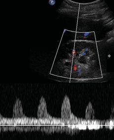
ultrasound High Resistive Index Peripheral Arterial Flow - ALiEM
aliem.com
source
Comments
ultrasound High Resistive Index Peripheral Arterial Flow | ALiEM
Upper Tract (Pelvicalyceal System and Ureter) | Radiology Key
Color Doppler imaging. The white arrow illustrates the perforation ...
Transesophageal echocardiographic apical four-chamber view of the heart ...
Third trimester of pregnancy. Two-dimensional echocardiography, the ...
Echocardiography showing pericardial effusion and thickened pericardium ...
Transesophageal echocardiography Transesophageal echocardiography ...
📃 Triploidy
Impact of the augmentation of the Impella flow level on mean arterial ...
Perioperative TEE (mid-esophageal bicaval view) immediately after ...
Doppler of umbilical artery showing normalization of systolic/diastolic ...
Continuous Doppler (CW). Apical 4 chambers section with the Doppler CW ...
A: mid-esophageal four chamber view. Left ventricle is enlarged with ...
Final Repositioned Portico (Abbott) Transcatheter Aortic Valve ...
Two-dimensional TTE oriented in a subcostal view with pulsed-wave ...
Transthoracic echocardiography shows left ventricular retraction due to ...
High-intensity transient signals (HITS) caused by microbubbles in the ...
transplant renal vein thrombosis ultrasound waveform - Google Search ...
ASD Rims by TTE and TEE: I | Download Scientific Diagram
Echocardiography before LDLLT. The right ventricle is dilated and ...
Typical morphological appearance of the Doppler signal "dagger−shaped ...
Summary of reported cases of Brevundimonas vesicularis infection in ...
Medial early diastolic mitral annular velocity (e′) measured by ...
Intraoperative transesophageal echocardiography in the midesophageal ...
Agitated saline bubble study during intracardiac echocardiography ...
File:Prolaps of AMVL and PMVL E00585 (CardioNetworks ECHOpedia).webm ...
M mode echocardiography showing paradoxical systolic and diastolic ...
Schematic diagram showing the aortic arch anatomy. The usual anatomy of ...
M-mode echocardiogram of left ventricle in the region of papillary ...
High speed at TIPS level | Download Scientific Diagram
Representative pulsed Doppler image showing the PEP and LVET time ...
An example of transcranial Doppler ultrasound with the agitated saline ...
FIGURE E Echocardiogram of the patient. The echocardiogram showed ...
Transthoracic echocardiography showing acute diastolic dysfunction ...
Parasternal long-axis view after thrombolysis showing a large mass ...
Doppler-Equation-Ultrasound
Liver-Doppler-Ultrasound
Thermal-Index-Ultrasound
Hepatic-Vein-Doppler-Ultrasound
Color-Doppler-Ultrasound
Normal-IVC-Waveform-Ultrasound
High-Resistance-Waveform-Ultrasound
Doppler-Ultrasonography
Renal-Doppler-Ultrasound
Low-Resistance-Waveform-Ultrasound
Transcranial-Doppler-Ultrasound
Renal-Artery-Stenosis-Doppler-Ultrasound
Umbilical-Cord-Doppler-Ultrasound
Renal-Arteries-Ultrasound
Pulsatility-Index-Ultrasound
Edv-Ultrasound


.webm/120px--Prolaps_of_AMVL_and_PMVL_E00585_(CardioNetworks_ECHOpedia).webm.jpg)


