Please enter url.
Login
Logout
Please enter url.
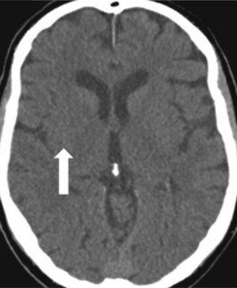
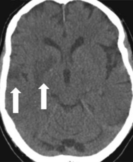
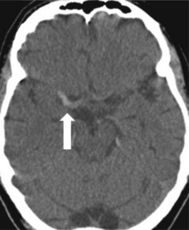
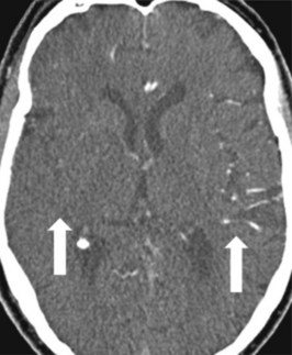
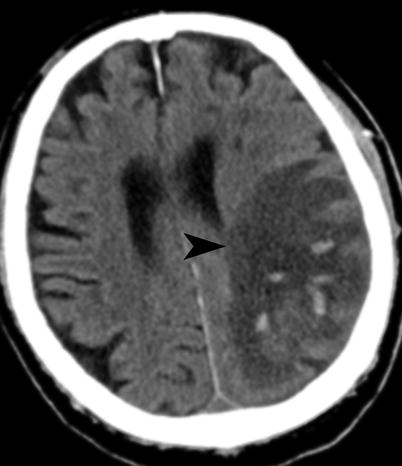
A 68-year-old woman with a left hemiplegia following a conscious ...
basicmedicalkey.com
source
Comments
A 68-year-old woman with a left hemiplegia following a conscious ...
A 68-year-old woman with a left hemiplegia following a conscious ...
A 68-year-old woman with a left hemiplegia following a conscious ...
MRI changes associated with acute status epilepticus. A middle-aged man ...
Clippers Syndrome Radiology - Kelis Guthrie
Post-intravenous contrast CT brain scan demonstrating right cerebral ...
The soft-tissue-window of a cerebral CT of a young patient demonstrates ...
(PDF) Case 14694 Severe hypoxic-ischemic brain injury: the reversal ...
CT brain with a focus of air in the cerebral venous system in the right ...
Cerebrovascular Accident in a Pediatric Patient Presenting With ...
Optimal window settings. (a) Standard window setting for soft tissue or ...
At 3 months of age, T1-weighted magnetic resonance imaging showed a ...
Category:CT images of cerebral infarction - Wikimedia Commons
Multifocal subdural haematoma of the tentorium cerebelli (indicated by ...
EURORAD - Radiologic Teaching Files
Ventriculo peritoneal shunt placement to treat hydrocephalus ...
The porencephalic cyst directly communicates with the posterior horn of ...
emDOCs.net – Emergency Medicine EducationUnusual Stroke Presentations ...
abnormal mri brain Ependymitis - Radiology Imaging
Figure1.Chest CT from a previous institution. A cavitary nodule was ...
-CT of the head revealed a hyper-dense sign in the right middle ...
CT scan of the brain without contrast showing an infarct in the left ...
Stroke and Its Imaging Evaluation | Radiology Key
(Case 2) Axial brain MRI indicating a lesion in the right frontal lobe ...
Computed tomography scan of the brain showing complete resolution of ...
(a) Preoperative CT scan showing enlarged ventricular system. (b) CT ...
Bilateral medial thalamic infarcts, a typical finding due to occlusion ...
Axial noncontrast head CT. Marked dilatation of lateral ventricles with ...
Bilateral medial thalamic infarcts, a typical finding due to occlusion ...
-Axial noncontrast CT soft tissue window (a) at the level of skull base ...
Cerebral oedema and dural sinus thrombosis in an adolescent with ...
Internet Scientific Publications
Sagittal section of brain CT showing significant pneumocephalus with ...
Second CT scan brain without contrast showing clearly visible ...
Axial brain nonenhanced CT scan in parenchymal window showing two ...
Monoplegia
Diplegia
Hemiparesis-vs-Hemiplegia
Hemiplegic-Cerebral-Palsy
Spastic-Diplegia
Ataxic-Hemiparesis
Lower-Limb-Spasticity
Left-Hemiparesis
Hemineglect
Hemiplegic-Gait
Spastic-Arm
Paraparesis
Paraplegia-Hemiplegia-Quadriplegia
Spastic-Diplegic-Cerebral-Palsy
Hemiplegia-Face
Cerebral-Infarction