Please enter url.
Login
Logout
Please enter url.
Carotid Artery Stenosis Chart Ultrasound
mavink.com
source
Comments
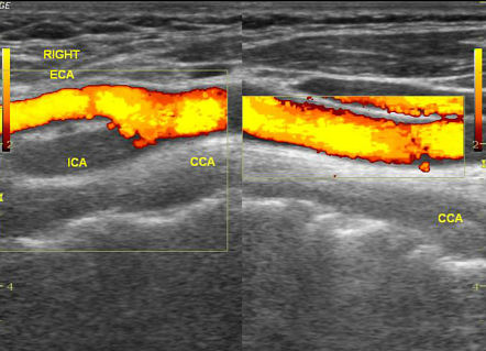
Carotid Artery Stenosis Ultrasound
Carotid duplex scanning showing significant atherosclerotic plaque ...
Fluoroscopic Image of Occlusive Thrombus Within the Axillary Artery ...
Bronchial atresia in an 18-year-old girl being evaluated for fever. CT ...
Image | Radiopaedia.org
Steal syndrome in a brachiocephalic fistula with distal hypoperfusion ...
Ultrasound cross-sectional image of the thoracic paravertebral space ...
Transverse view of the left carotid arteries. a. in the middle third of ...
a Complete tear of the distal biceps tendon (arrow). b Retracted ...
(PDF) Detection of anatomical variation during left internal jugular ...
(PDF) Ultrasound-Guided Cervical Nerve Root Block
The digastric muscle posterior belly (DMPB) and hypoglossal nerve ...
Figure 4:Color Duplex Assessment of 4th and 5th Internal Mammary Artery ...
(a) Ultrasound-guided C2 transverse process block (left side). (b) The ...
Carotid duplex revealing resolution of stenosis. | Download Scientific ...
Bilateral comparative examination of a thickened, hypoechogenic ...
Ultrasound-measured jugular venous pressure imaged in a semi-recumbent ...
The 1 × 1 cm defect in the left scrotum with purulent discharge found ...
Perforator of Duplex ultrasound image: a perforator arising from the ...
(PDF) An uncommon cause of anterior elbow pain: Diagnosis and injection ...
A and B) Ultrasound shows solid, well-defined, hypoechoic mass located ...
Axial images of trachea and pretracheal structures on ultrasound ...
A) Ultrasound (US) doppler images showing a pseudoaneurysm originating ...
Carotid duplex ultrasonography conducted on the eleventh day of ...
The different thickness of the intestinal wall of the appendix orifice ...
CTA demonstrating complete occlusion of the proximal right popliteal ...
Left, Preoperative contrast-enhanced duplex scan of the internal ...
(PDF) Routine preoperative doppler ultrasound examination of arterial ...
Transverse scan of left upper neck showing the anatomical relationship ...
Vascular Laboratory Testing | Clinical Gate
Icd 10 Code For Calcific Tendinitis Left Hip
Ultrasound imaging of the posterior-inferior tibiofibular ligament ...
Brachial plexus echography performed with ultrasound transducer placed ...
-A) Transverse section of the right common carotid artery with color ...
A, First anastomosis of a sequential LIMA-diagonal-LAD: side-to-side ...
Carotid-Artery-Scan
Abnormal-Carotid-Doppler
Carotid-Artery-Blockage
Carotid-Artery-Test
Carotid-Artery-Plaque
Carotid-Artery-Symptoms
Carotid-US
Carotid-Artery-Thrombosis-Ultrasound
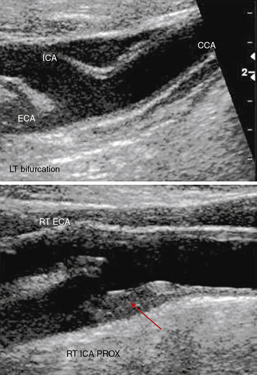
Carotid-Artery-Bifurcation
Normal-Carotid-Ultrasound
Carotid-Artery-Occlusion
Carotid-Bruit
Subclavian-Artery-Ultrasound
Carotid-Artery-Disease
Carotid-Artery-Neck
Carotid-Artery-On-Thyroid-Ultrasound