Please enter url.
Login
Logout
Please enter url.
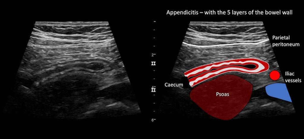
Appendix Ultrasound Normal Measurements
mavink.com
source
Comments
Ultrasound Case 083 • LITFL • POCUS Self-Assessment Quiz
Ultrasound and Mapping of Neck Lymph Nodes | Radiology Key
scrotal ultrasound - Startradiology
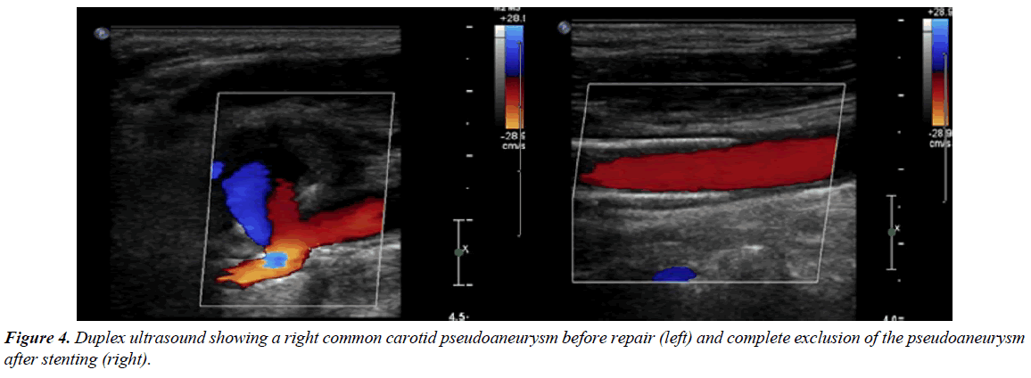
Endovascular treatment of a carotid pseudoaneurysm with covered stents ...
Case 2 ultrasound finding. a first-trimester ultrasound scan shows ...
Scout scan of the left femoral nerve (short axis) at the level of ...
Longitudinal colour Doppler ultrasound image at the level of the right ...
CTA demonstrating complete occlusion of the proximal right popliteal ...
Acute extensive right lower extremity edema and phlegmasia cerulea ...
Patient 1; Ultrasound Image of the Greater Occipital Nerve in Relation ...
Abdominal MRI An abdominal MRI showing a focal hepatic lesion (white ...
Ultrasound diffuse liver disease all things fibrosis,cirrhosis,us ...
Use of colour Doppler and M-mode ultrasonography to confirm the ...
PP-353 The Comparison of Isolated or Combined Failure on V.Saphena ...
Ultrasound strain imaging in assessment of false vocal folds in adults ...
True Radial Artery Aneurysm: Diagnosis and Treatment - Journal of ...
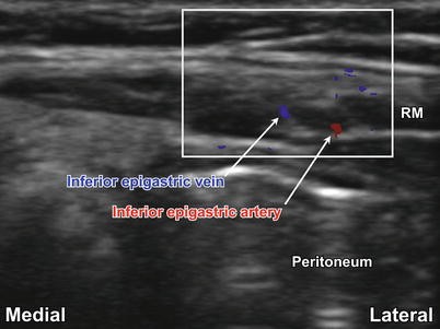
Rectus Sheath and Transversus Abdominis Plane (TAP) Blocks | SpringerLink
(PDF) IJV Phlebectasia: an approach algorithm
Left peroneal vein deep vein thrombosis (yellow arrow), axial view ...
Ultrasound in Clearwater, Dunedin and Palm Harbor
A) Transverse scan ultrasound of supraspinatus tendon showing ...
Figure1.Ultrasonography images (color flow Doppler imaging). A: A ...
Color Doppler image of common femoral vein. Spectral analysis depicting ...
Traumatic Superficial Subcutaneous Testicular Dislocation | Eurorad
Ultrasound image 18 h after block Figure 1: Ultrasound image 3 h after ...
The distension of the right CCA by vascular ultrasound measurements in ...
Anatomy location of bladder, vaginal canal, cervix and uterus. CT and ...
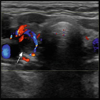
Ultrasound as a Localization Technique in Hyperparathyroidism ...
Cricoid cartilage and tracheal cartilage in longitudinal plane is seen ...
Ultrasound shoulder and knee joints | PPT
Situs inversus | Image | Radiopaedia.org
RadiologySpirit: Another sign to identify the ECA (external carotid ...
94 ideas de Ultrasonido en 2021 | ultrasonido, radiología, imagenologia
External carotid compression: a novel technique to improve cerebral ...
-A 28-year-old male with complete hypoechoic thrombosis in the right ...