Please enter url.
Login
Logout
Please enter url.
Microsurgical resection of endolymphatic sac tumors - Operative ...
optecoto.com
source
Comments
Temporal Bone Tumors | Radiology Key
The Inner Ear and Otodystrophies | Radiology Key
Spectrum of Third Window Abnormalities: Semicircular Canal Dehiscence ...
The Many Faces of Facial Nerve Schwannoma | American Journal of ...
Imaging of Temporal Bone Tumors | Radiology Key
Congenital malformations of the external and middle ear - European ...
Standardization of Temporal Bone CT Planes across a Multisite Academic ...
Non-Syndromic Sensorineural Hearing Loss in Children - Neuroimaging Clinics
Role of HRCT and MRI of the Temporal Bone in Predicting and Grading the ...
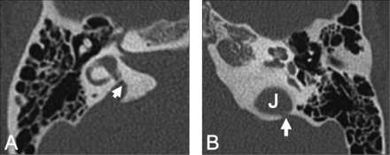
Range of abnormalities of the vestibular system seen in axial CT ...
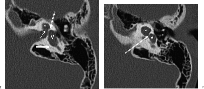
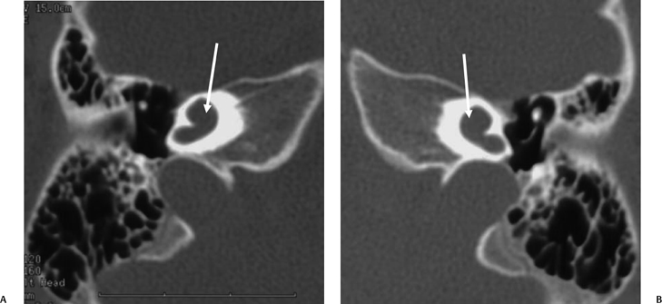
Imaging and clinical findings in large endolymphatic duct and sac ...
Infranuclear Facial Palsy: Importance of Imaging the Genicul ...
Impact of Imaging in Management of Otosclerosis - Otolaryngologic ...
The Inner Ear and Otodystrophies | Radiology Key
Figure 1 from Tympanic Plate Fractures in Temporal Bone Trauma ...
The Inner Ear and Otodystrophies | Radiology Key
(PDF) Endoscopic-assisted cochlear implant procedure in CHARGE syndrome ...
Spatial association between the cochleariform process and the tympanic ...
(PDF) Pneumatization of Mastoid Air Cells, Temporal Bone, Ethmoid and ...
Figure .. Lateral semicircular canal complex. (A) Axial temporal bone ...
Imaging Findings in Auto-Atticotomy | American Journal of Neuroradiology
Figure 1 from Imaging Case of the Month Charcot-Marie-Tooth Disease as ...
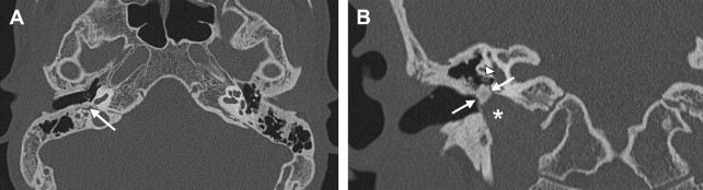
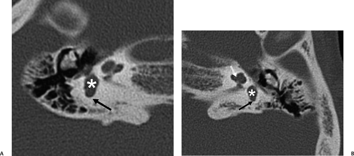
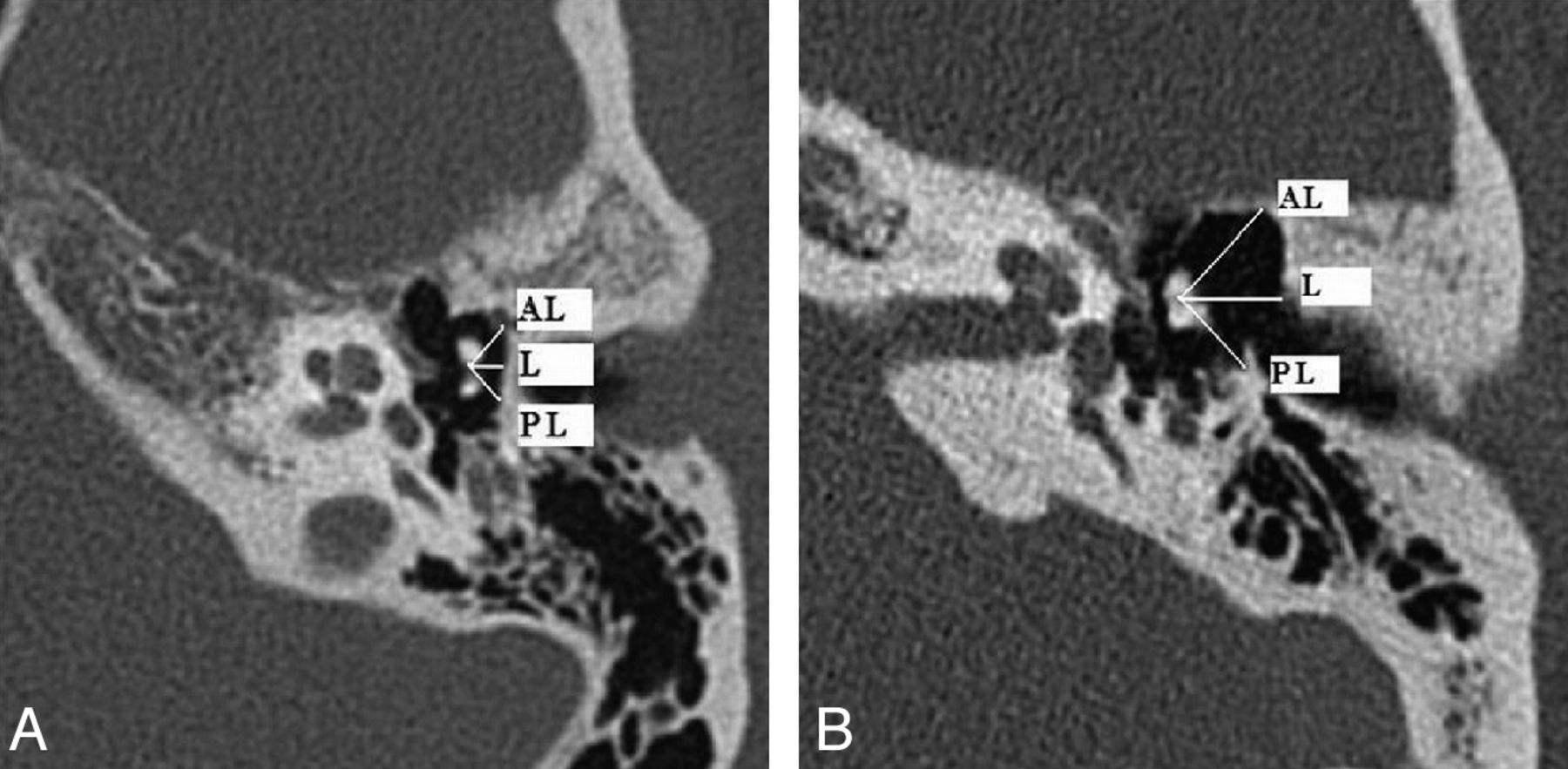
The vestibular aqueduct as seen on axial (A), coronal (B), sagittal ...
Computed tomography imaging: computed tomography showing the left ear ...
(A and B) Axial temporal bone computed tomography (CT) clearly showed ...
Imaging of Tinnitus - Neuroimaging Clinics
(A) Otoscopy showed a dark-red mass in the tympanic cavity. (B) Two ...
Imaging of post-traumatic hearing loss | Radiología (English Edition)
Cochlear Implantation: Systematic Approach to Preoperative Radiologic ...
| CT scan demonstrating (A) HSC-FND on coronal imaging, (B) normal HSC ...
5 Axial HRCT images of the right (a) and left (b) temporal bone in a ...
Congenital malformations of the external and middle ear - European ...
(PDF) Otosclerosis and complications of stapedectomy: CT and MRI ...
Barotrauma Presenting as Temporal Lobe Injury Secondary to Temporal ...
Otosclerosis and Dysplasias of the Temporal Bone - Neuroimaging Clinics
Endolymphatic-Sac-Tumor
Endolymphatic-Sac-Anatomy
Endolymphatic-Shunt
Endolymph
Endolymphatic-Hydrops
Endolymphatic-System
Ear-Sac
Endolymphatic-Mastoid-Shunt
Endolymphatic-Fluid
Inner-Ear-Sack
Endolymphatic-Appendage
Endolymphatic-Shunt-Austin
Vestibular-Sacs
Large-Endolymphatic-Sac-Anomaly-Radiology
Perilymph
Secondary-Endolymphatic-Hydrops