Please enter url.
Login
Logout
Please enter url.
Carotid Ultrasound Waveforms
mungfali.com
source
Comments
Figure 7 from A spectrum of Doppler waveforms in the carotid and ...
Diagram of the blood supply to the pancreas, stomach and spleen ...
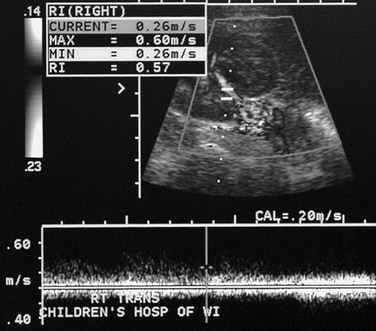
(PDF) Doppler ultrasound studies in renal arteries of normal newborn babies
ACI stenosis (80%) with a mixed, predominantly fatty plaque. | Download ...
Ultrasound images of Inflammatory bowel conditions - Radiology Imaging
(PDF) Mumps epididymo-orchitis: Sonography and color Doppler ...
Cervical artery dissection: dissection of ACC and ACI ∑ two lumina ...
(PDF) Parvus tardus waveform suggesting renal artery stenosis ...
Renal Vein Doppler Sonography of Obstructive Uropathy | AJR
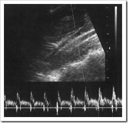
Normal range for resistance index (RI) of umbilical artery flow ...
US of Neurovascular Occlusive Disease: Interpretive Pearls and Pitfalls ...
Bevel of needle is in the lumen of vein | Download Scientific Diagram
The intima/media complex is depicted in panel A for a normal brachial ...
Continuous wave Doppler. Two crystals are used - one transmitting ...
Peak systolic velocity, end diastolic velocity, and resistive index ...
An example of normal respiratory phasicity of venous fl ow as the ...
B-mode ultrasound image of the radial artery. The arterial lumen is ...
Example of hemodynamically significant stenosis of the left renal ...
Sonogram of SDFT allograft at 90 days postoperatively revealed complete ...
Peak systolic velocity, end diastolic velocity, and resistive index ...
[PDF] A spectrum of Doppler waveforms in the carotid and vertebral ...
(PDF) B-scan ultrasonography of ocular abnormalities: A review of 182 dogs
Common Errors in the Doppler Ultrasound Display of Uterine Blood Flow ...
(PDF) Serendipitous diagnosis of aortic coarctation by bilateral parvus ...
Hypertension and aortic coarctation in an asymptomatic teenage girl ...
Colour Doppler ultrasound showing flow in the ophthalmic artery and ...
Colored coded duplex Doppler in a patient with liver cirrhosis and ...
Annotated version of Figure 21. Fibromatosis colli. Ultrasound. Right ...
(PDF) Evaluation of ultrasound for central venous access in ICU by an ...
Doppler breast ultrasound demonstrates hypo-echoic solid right breast ...
Vascular Conditions | Radiology Key
Subclavian steal syndrome: vertebral artery ∑ inversed spec- trum due ...
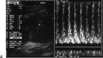
Normal umbilical artery flow velocity waveform at 48 days’ gestation ...
Hypoechoic, solid nodule with smooth borders. Although there is no halo ...
Ultrasound image showing numerous, small, echogenic foci of ...
Vascular-Doppler-Ultrasound
Continuous-Wave-Doppler-Ultrasound
Arterial-Doppler-Ultrasound
Types-of-Doppler-Ultrasound
Portal-Vein-Doppler-Ultrasound
Color-Flow-Doppler-Ultrasound
Renal-Artery-Doppler-Ultrasound
Carotid-Doppler-Ultrasound
Lower-Extremity-Arterial-Doppler-Ultrasound
Biphasic-Waveforms-Arterial-Doppler
Hepatic-Vein-Doppler
Aortic-Stenosis-Waveform
Umbilical-Artery-Doppler-Ultrasound
Spectral-Doppler-Ultrasound
Pulse-Wave-Doppler-Ultrasound
Testicular-Doppler-Ultrasound


![[PDF] A spectrum of Doppler waveforms in the carotid and vertebral ...](https://d3i71xaburhd42.cloudfront.net/981fb6c7f30a38744a94b7e362331b02599042e4/250px/3-Figure3-1.png)