Please enter url.
Login
Logout
Please enter url.
Nadia Pinheiro on LinkedIn: Echo showed marked concentric LVH with ...
linkedin.com
source
Comments
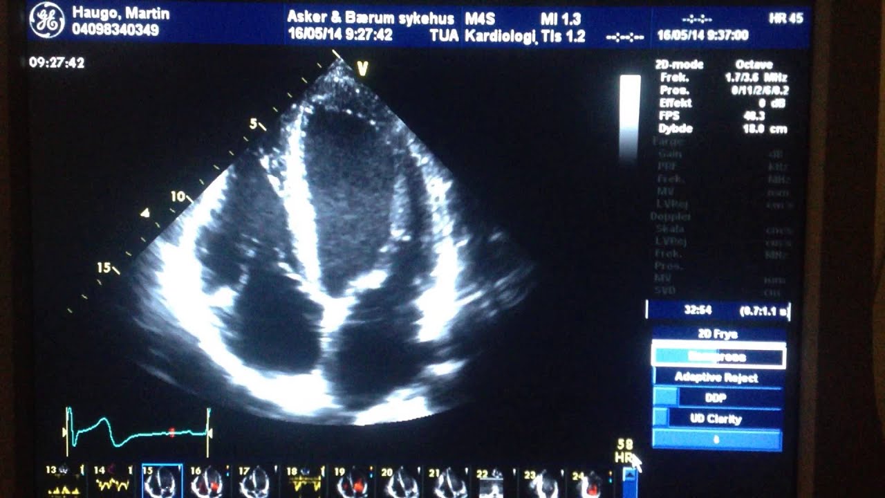
Ultralyd hjerte - YouTube
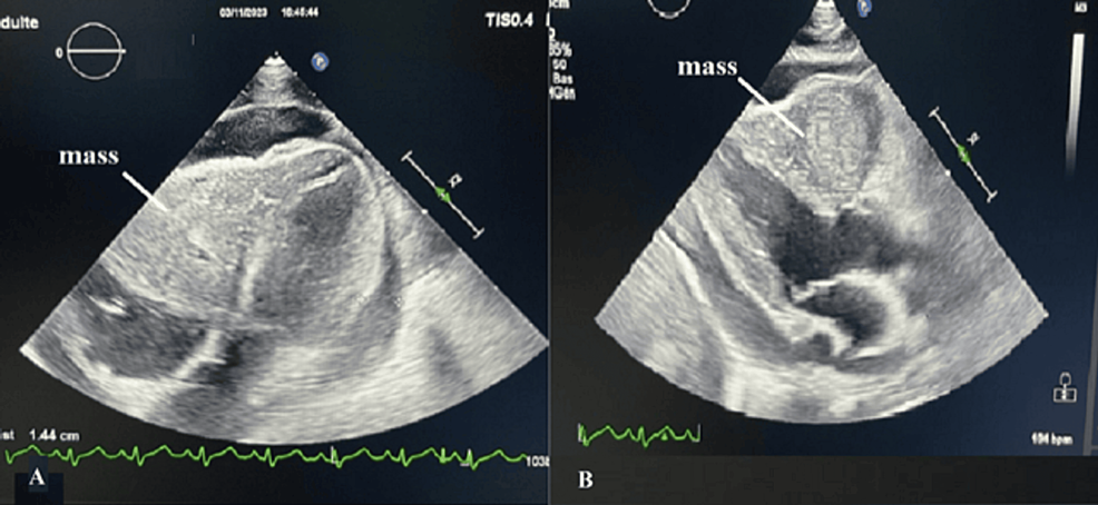
Cureus | A Giant Primary Angiosarcoma Invading the Right Heart in a ...
Aortic valve prolapse - YouTube
Replacement of the Ascending Aorta With Aortic Root Remodeling Without ...
Technology
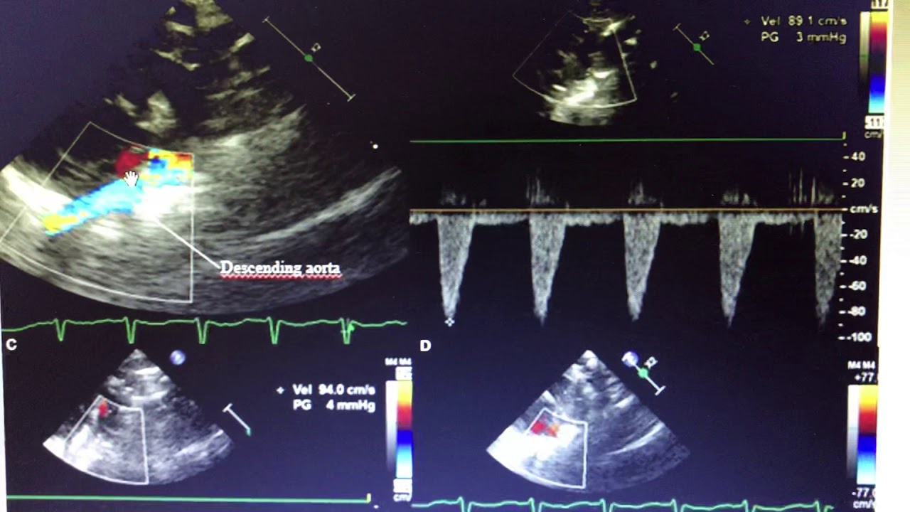
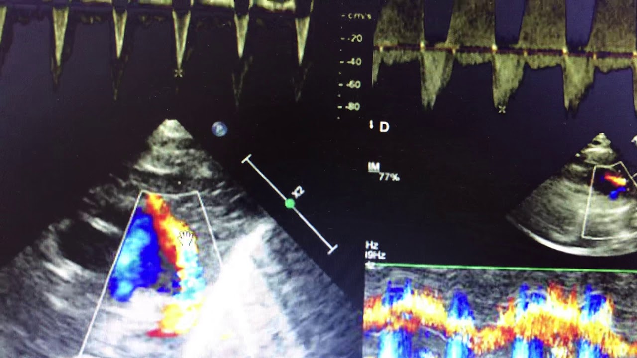
Descending aortic Doppler in PDA - YouTube
Acute aortic dissection
Biatrial dilatation // RCMP - YouTube
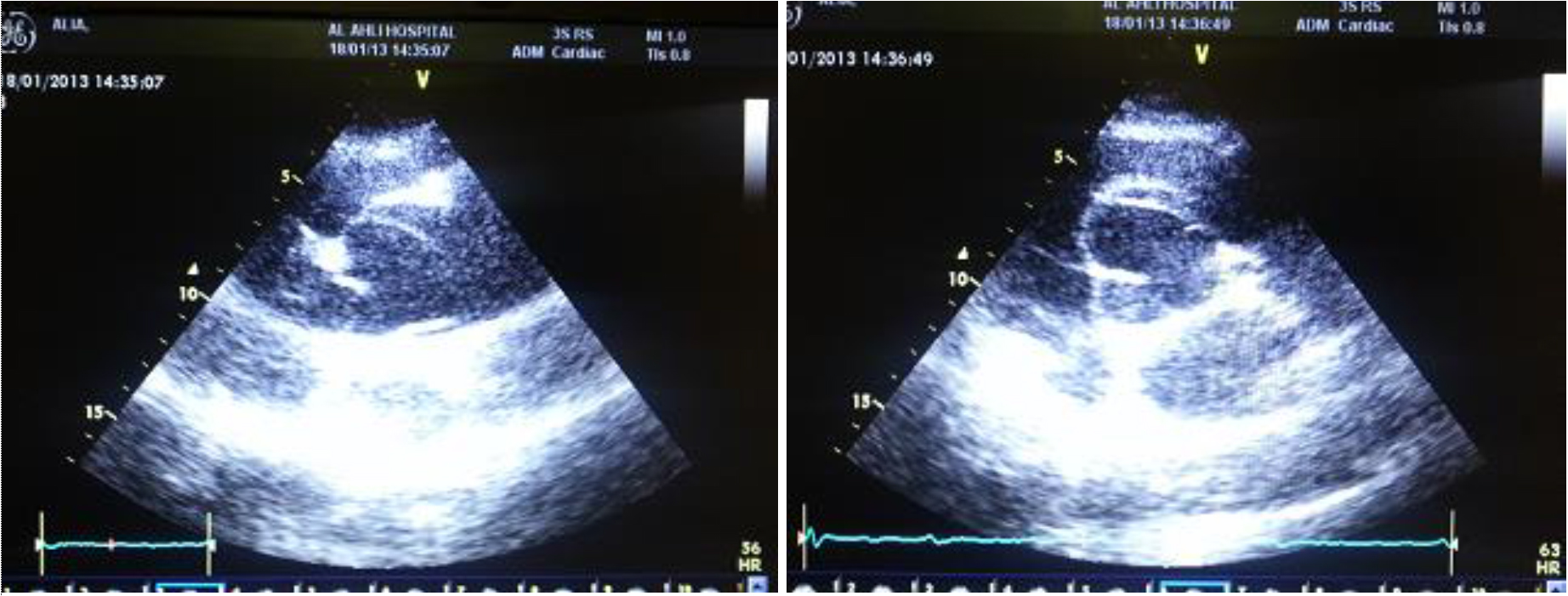
Echocardiogram - Aortic aneurysm - The largest aortic root - YouTube
TTE with color Doppler showing the atrial septal defect. | Download ...
2d echocardiogram- Vegetation on ICD pacer wire - YouTube
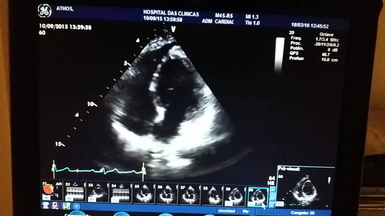
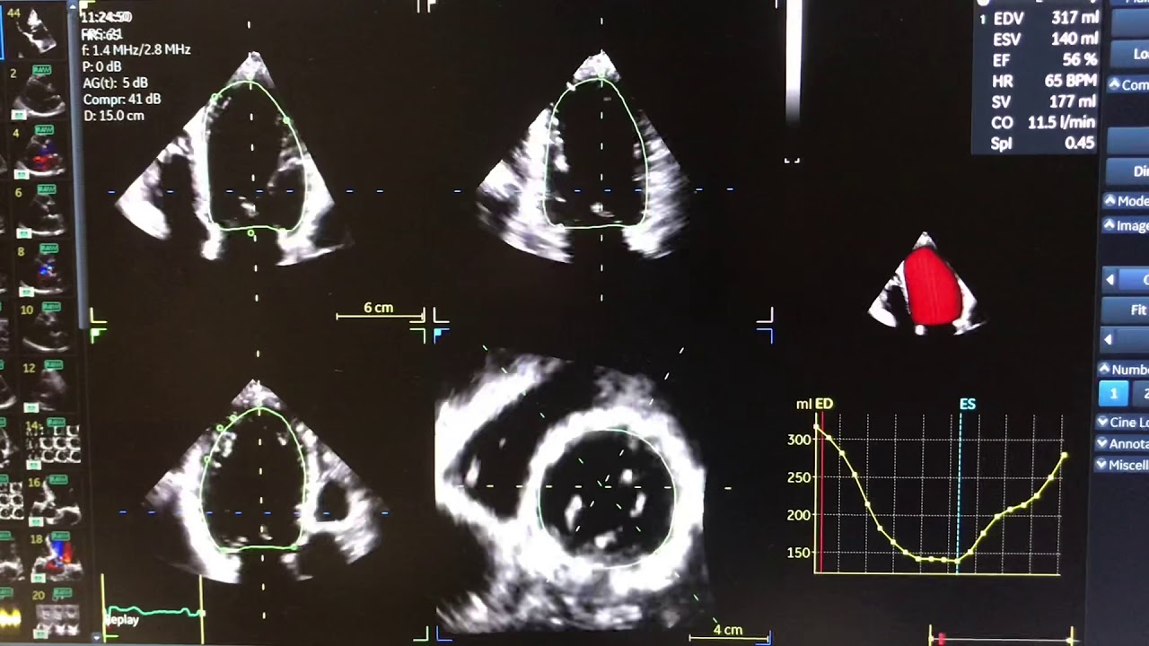
Can you visually estimate ejection fraction in this echo? Previous one ...
Transthoracic echocardiography in the parasternal long axis window ...
Echocardiogram - Aortic dissection with severe aortic regurgitation ...
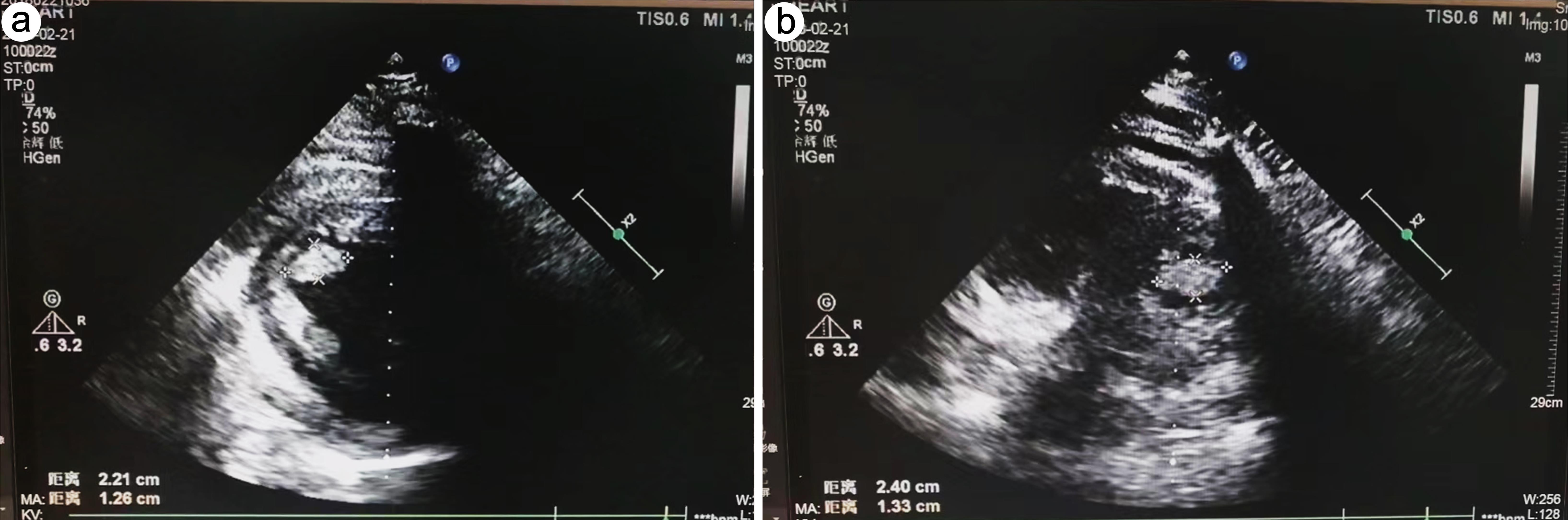
Images of the transthoracic echocardiogram showing thickened ...
Cardiac Tamponade on Cardiac Echo Apical 4 Chamber View - YouTube
Pattern, Precipitants and Short Term Outcome of Heart Failure Patients ...
First echocardiography of patient in hospital. | Download Scientific ...
OPTOMISE 2D in ultrasound philips sonos 7500 part 4 - YouTube
A VSD occluder was advanced along the sheath. When the left to right ...
Cardiac2 - YouTube
ECHO STRESS.MOV - YouTube
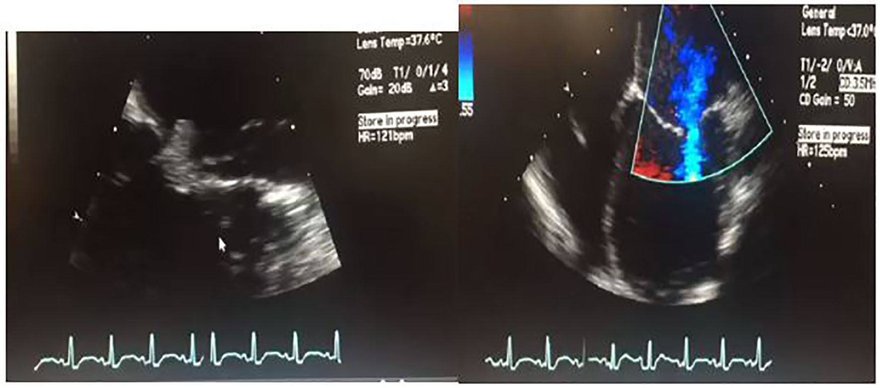
Transesophageal echocardiography showed a floating and vibratile ...
Atrial septal aneurysm - YouTube
Libman-Sacks endocarditis - wikidoc
PDA with left to right shunt - YouTube
Mitral regurgitation 1 - YouTube
20190828_04_Mitral stenosis and regurgitation_Dr Arupratan Maiti - YouTube
2 D echocardiogram Changes in PAH - YouTube
Treatment of the Left Ventricular Thrombus with Integrated Traditional ...
Aneurisma de ventrículo esquerdo - YouTube
2D echo - Severe MR, giant LA pericardial effusion - YouTube
Belhassen anterior fascicular ventricular tachycardia: a case in a ...
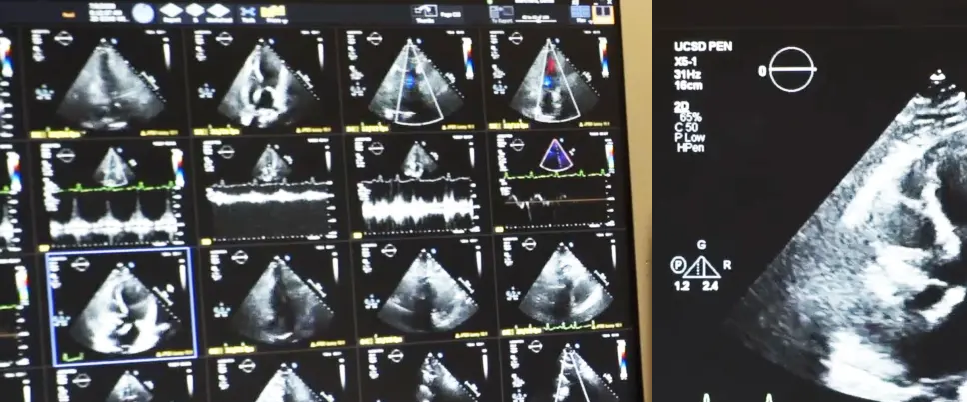
Cardiac Imaging


-short%20axis%20view%20(B)%20Long%20axis%20view_update.jpg)