Please enter url.
Login
Logout
Please enter url.
Head MR and classic cases | Radiology Key
radiologykey.com
source
Comments
Brain MR imaging in a control neonate; comparison between the perfusion ...
Figure 1:Isolated Shoulder Weakness due to a Small Cortical Infarction ...
Axial CT scan of brain without contrast. Small lacunar infarct ...
Pelizaeus-Merzbacher Disease: Background, Etiology, Epidemiology
Diagnostic testing in prion disease. | Download Scientific Diagram
Ring-enhancing lesions: axial T2-weighted image showing a rim lesion ...
Brain MRI revealed increased DWI and T2 signal in the caudate nuclei ...
MRI brain shows diffuse high signal intensity involving subcortical ...
Cureus | Changes in the Brain Connectome Following Repetitive ...
2 Free-floating Thrombus of the Left Internal Carotid Artery | Neupsy Key
Pathology of the left supraclavicular lymph node shows a poorly ...
-Diffusion weighted images (DWI), ADC maps and axial T2-FLAIR weighted ...
The degree of postictal hypoperfusion is directly related to seizure ...
Frontiers | Low-Field MRI: How Low Can We Go? A Fresh View on an Old ...
Basal Ganglia | Brain
Neuroimaging Methods in the Study of Childhood Psychiatric Disorders ...
Dr Balaji Anvekar's Neuroradiology Cases: Hyperdense thalami ...
Coronavirus Disease: Subacute to Chronic Neuroimaging Findings ...
Laminar cortical infarcts with hemorrhagic transformation. (a) T2 ...
Normal diffusion magnetic resonance imaging examination findings on the ...
The ‘windows of opportunity’ for treatment optimization in MS. Early ...
Coincident VZV Encephalitis and Otomastoiditis EMRA
Diffusion-weighted magnetic resonance images obtained after ...
MRI findings of striatocapsular-region infarction. A: Initial DWI image ...
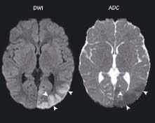
(a and b) Diffusion-weighted imaging and apparent diffusion coefficient ...
Punctate white matter lesion (PWML): axial T1W (A), axial T2W (B) and ...
2 Free-floating Thrombus of the Left Internal Carotid Artery | Neupsy Key
Diffusion Tensor Imaging
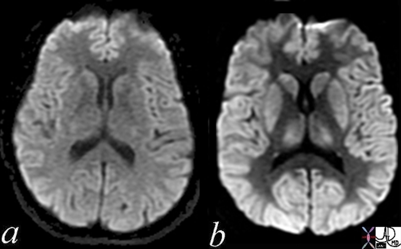
Axial Brain MRI in DWI sequence. Panels (a) and (b) show diffusion ...
A. An axial fractional anisotropy and B. mean diffusivity map (mm 2 s ...
diffuse axonal injury
ROIs used in the normal-appearing gray and white matter and ...
Symptomatic Narcolepsy or Hypersomnia, with and Without Hypocretin ...
Persistent Choreoathetosis in a Fatal Olanzapine Overdose: Drug ...
(PDF) MRI Abnormalities Induced by Seizures