Please enter url.
Login
Logout
Please enter url.
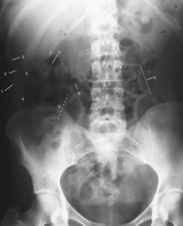
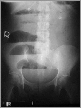
Abdominal X-Ray: Indications and Normal Findings
urology-textbook.com
source
Comments
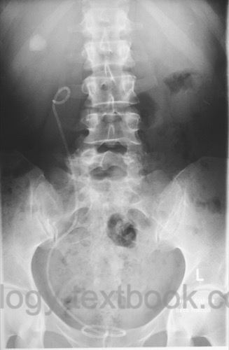
Plain abdominal radiograph with sleeve catheter in situ. The 6-cm-long ...
Copy and Scanning - ADV Photo
The Urinary Tract in Pregnancy | Plastic Surgery Key
The abdomen | Radiology Key
Calculus-freeafter ureteroscopy. doi:10.1371/journal.pone.0087634.g005 ...
Intravenous Urography: Technique and Interpretation | RadioGraphics
Periaortitis with Ureteral Obstruction After Endovascular Repair of an ...
Kidney, ureter, and bladder X-ray postoperatively | Download Scientific ...
Staghorn calculi (coral calculi) | Radiology Case | Radiopaedia.org ...
Urography after 60 months. | Download Scientific Diagram
Image Library Bladder Infection | BMC Radiology
staghorn_calculus_struvite_stone_ammonium_magnesium_phosphate_proteus ...
JaypeeDigital | eBook Reader
نماذج اسئلة سلايدات pptx - جراحة - Muhadharaty
GI Flashcards | Quizlet
Plain film of liver transplant recipient showing Silastic biliary stent ...
Spectrum of congenital renal anomalies presenting in adulthood ...
-Intravenous urography. Left kidney chronic pyelonephritis with altered ...
Acute Renal Colic from Ureteral Calculus | NEJM
Abdominal plain film showing the lost band within the small bowel ...
b: Cystolithotomy access obtained through a 10mm sheath by using the ...
Fecal impaction: a cause of isolated small bowel dilatation on ...
Case 22-1967 — Large Mass in the Left Upper Quadrant of the Abdomen | NEJM
Welcome to LearningRadiology
Image | Radiopaedia.org
A new cause of curvilinear renal calcification: calcified ...
The abdominal X-ray film revealed the course of the peritoneal catheter ...
Figure 1 from Adrenal myelolipoma with osseous metaplasia and ...
Intravenous Urography: Technique and Interpretation | RadioGraphics
Seminal Vesicle Cyst with Ipsilateral Renal Agenesis | AJR
22 Must Sees-2012
Image | Radiopaedia.org
Pelvic/abdominal X-ray showing osteopenia. | Download Scientific Diagram
Gallstones
Frontal radiograph of a 45-year-old woman with pseudotumor cerebri ...