Please enter url.
Login
Logout
Please enter url.
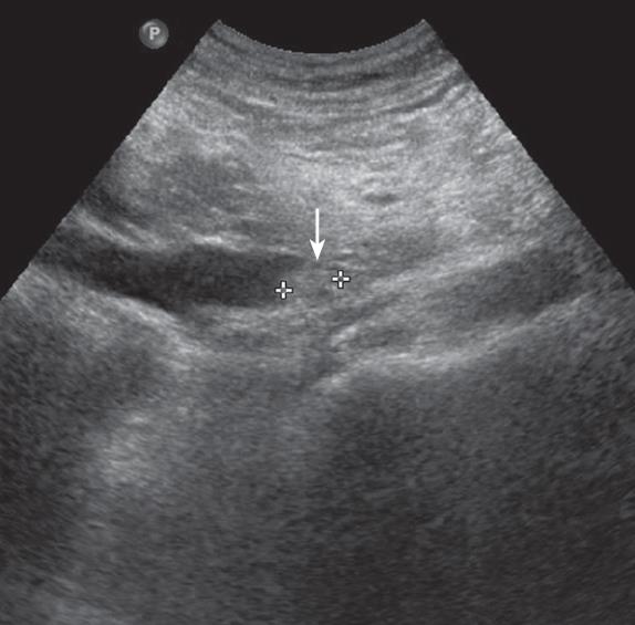
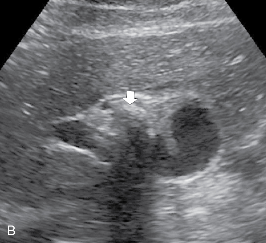
Cirrhosis Ultrasound
ar.inspiredpencil.com
source
Comments
Imaging of the cirrhotic liver | Radiology Key
Multimodality Imaging of Liver Infections: Differential Diagnosis and ...
Hepatic peliosis | Eurorad
pancreatitis ultrasonido - Ecosia | Ultrasonido y Radiografia
A 65-year-old man with degenerative hydatid cyst. Sonography reveals a ...
-Ultrasound-guided biopsy showing the needle (straight white arrow ...
Colonic diverticulitis: sonographic findings | Eurorad
Ectopic Pregnancy | Radiology Key
Differential Diagnosis of Intracranial Cystic Lesions at Head US ...
Non-invasive evaluation of liver cirrhosis using ultrasound - Clinical ...
Irregular, asymmetrical thickening of the GE wall (arrows) in a patient ...
Ecografía torácica | Archivos de Bronconeumología
Hepatocellular Carcinoma Treated with Radio-frequency Ablation ...
Vesicoureteral Reflux in Children with Urinary Tract Infection ...
A lung ultrasound of a 64-year-old male, showing lung consolidations ...
CT scan showing a retroperitoneal abscess (arrow) due to retained ...
From the Archives of the AFIP | RadioGraphics
DA16-DB1-Cystic_Dilation_of_Distal_Ureter-FFU2.gif | Abdominal Key
From the Archives of the AFIP | RadioGraphics
-Carcinoma vescicale avanzato. Tumore vegetante a larga base di ...
A 12-year-old female with complaints of vague upper abdominal ...
(PDF) Hepatosplenic Sarcoidosis: Contrast-Enhanced Ultrasound Findings ...
Cecal carcinoma. US reveals asymmetric, hypoechoic, circumferential ...
Carcinoma in situ arising in a tubulovillous adenoma of the distal ...
Histologic findings. (A) Histologic findings of liver reveals diffuse ...
Cholecystitis | Radiology Key
US of Gastrointestinal Tract Disease | RadioGraphics
Ultrasound-guided biopsy of a hypoechoic liver lesion: | Open-i
Hypoechoic Lesion In Liver In Hindi - andre
Langerhans cell histiocytosis in children under 12 months of age: The ...
Ultrasonographic image of an 8-month-old Thoroughbred filly (case 5 ...
Multiple renal masses of renal lymphoma. Axial contrast CT (a ...
WK 2 LIVER PATHOLOGY Ultrasound images on Metastases with bull’s-eye ...
Ultrasound and MDCT Findings in Cystadenocarcinoma of the Appendix with ...
A torsed ovary with perifollicular edema. This is a sagittal view with ...
Liver-Imaging
Cirrhotic-Liver-Ultrasound
Cirrhotic-Liver-CT
Cirrhotic-Liver-Morphology
Liver-Cirrhosis-MRI
Cirrhotic-Liver-Disease
Cirrhotic-Liver-Autopsy
Liver-Cirrhosis-Stages
Real-Cirrhotic-Liver
Cirrhotic-Liver-Specimen
Advanced-Liver-Cirrhosis
Dog-Cirrhotic-Liver
People-with-Cirrhosis-of-the-Liver
Normal-and-Cirrhotic-Liver
Cirrhotic-Liver-Image.jpg
Calcified-Cirrhotic-Liver


