Please enter url.
Login
Logout
Please enter url.
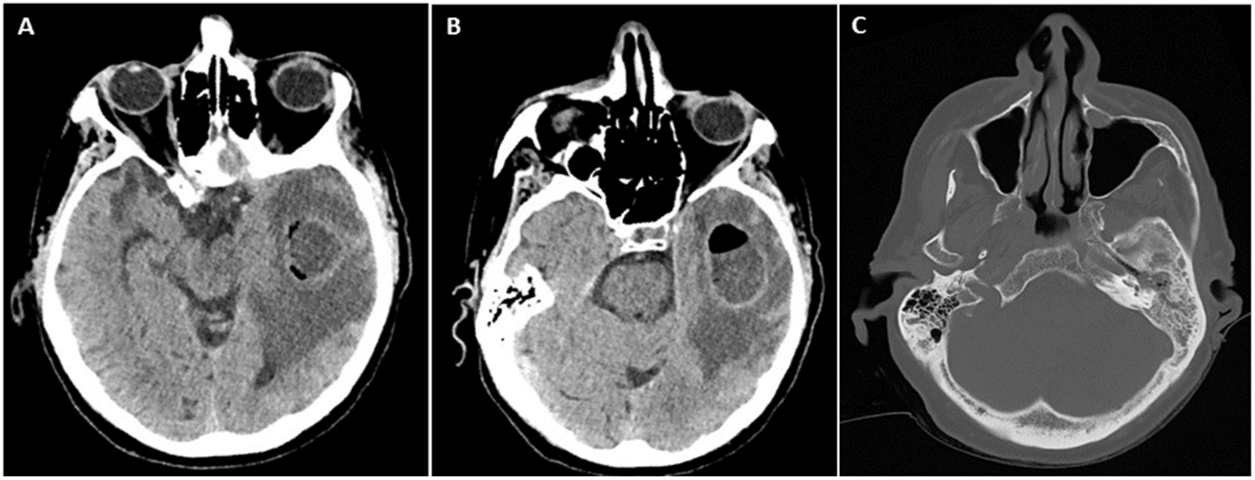
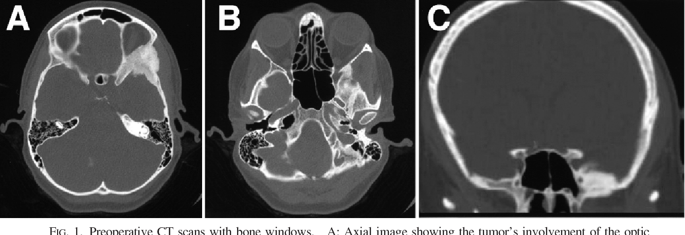
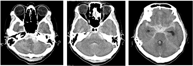
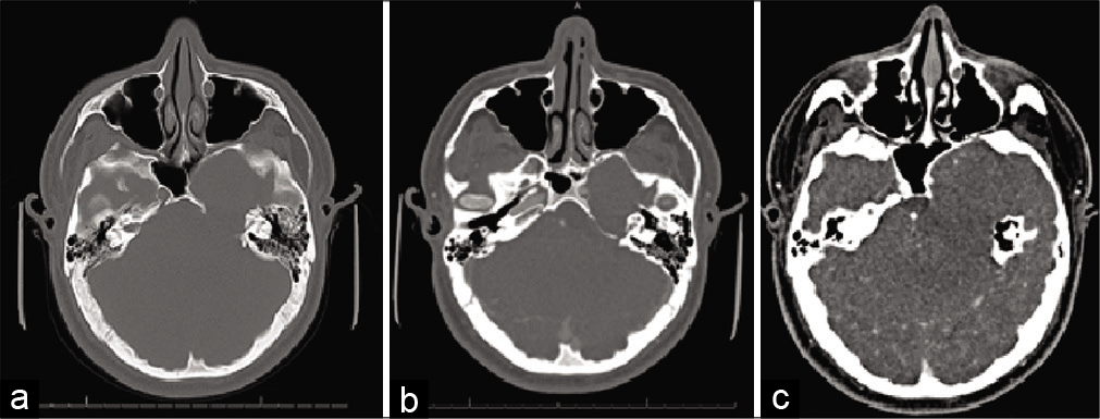
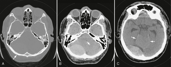
Axial slices of CT brain demonstrating (A) intraparenchymal haematoma ...
researchgate.net
source
Comments
Arterial thrombosis following first-dose ChAdOx1 vaccination: a case ...
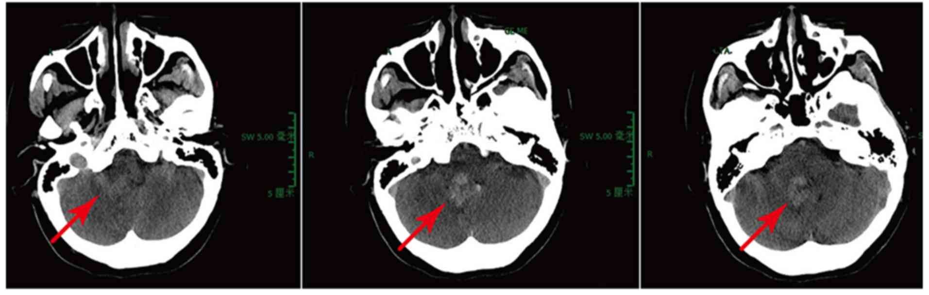
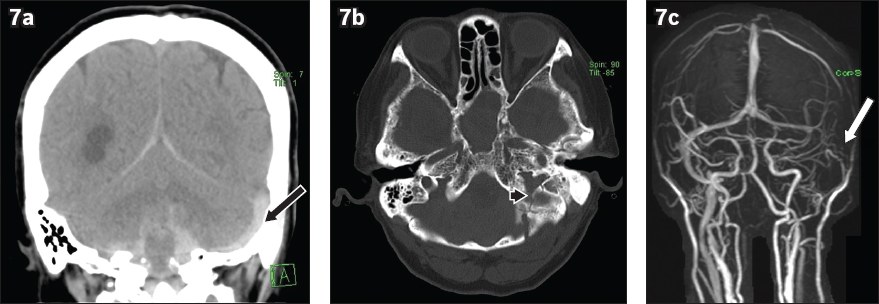
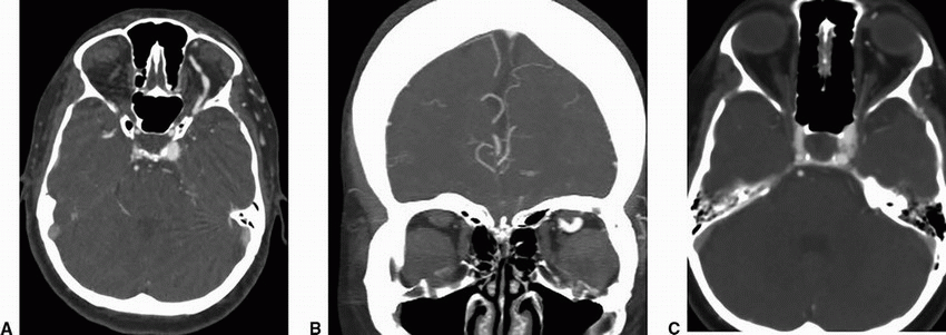
Case 2. A: Preoperative axial CT scan demonstrating left downward ...
Radiology MRI: Optic Disc Drusen
Surgical treatment of cerebellar infarction: long-term outcomes and ...
Is there a Connection Between COVID-19 and Vision Loss?
Hemorrhagic Dissection of the P1 Segment of Posterior Cerebral Artery ...
Bilateral Earlobe Crease (Frank’s Sign) and Multifocal Vascular Disease ...
Surgical Neurology International
Images of a cadaver head scanned on a pre-clinical hybrid dual-source ...
41 Epidermoids, Dermoids, and Other Cysts of the Skull Base | Neupsy Key
Cureus | Intracranial Hypotension Syndrome: The Importance of ...
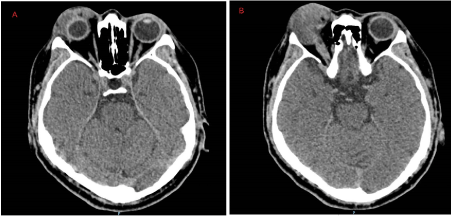
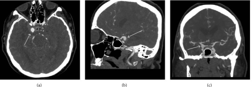
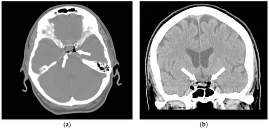
Preoperative CT-Angiogram. CT: computed tomography, CTA | Open-i
JCM | Free Full-Text | Air Embolism: Practical Tips for Prevention and ...
Fracture and Hemorrhage | Radiology Key
Vascular Abnormalities | Radiology Key
Cureus | A Method for Combining Thin and Thick Malleable Titanium Mesh ...
9 Carotid-Cavernous Fistulas | Neupsy Key
Radiology Case - Headache [clinical] : r/medicalschool
Treatment of a giant complicated distal posterior inferior cerebellar ...
Squamous Cell Carcinoma Arising from Sinonasal Inverted Papilloma ...
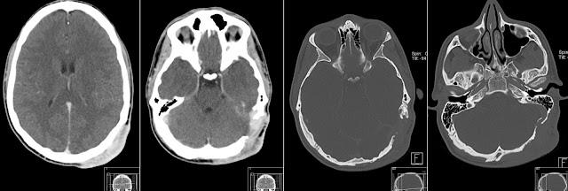
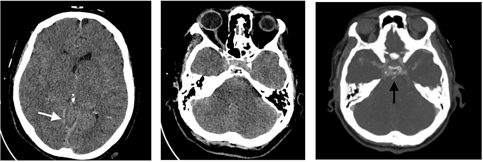
Initial brain CT shows. A: diffuse acute subarachnoid hemorrhage ...
Mastoiditis and Tegmen Tympani Defect Leading to Intracranial Abscess ...
Dense calcification in a GH-secreting pituitary macroadenoma in ...
Endovascular Management of Intracranial Dural AVFs: Principles ...
Peripheral orbital nerve schwannoma: Case report
Critical care for concomitant severe traumatic brain injury and acute ...
Radiology MRI: Neurofibroma
Cerebral venous thrombosis: a spectrum of imaging findings | SMJ
Cerebellar Hemangioblastoma - The American Journal of Medicine
Cerebellar Disease Mimicking Cerebrotendinous Xanthomatosis: Langerhans ...
Figure 1 from Management of bone-invasive, hyperostotic sphenoid wing ...
Immediate postoperative and post seizures CT head without contrast ...
Computed tomography images of a 35-year-old man with chordoma invading ...
The CT scan of the brain with contrast showing (A) before treatment ...
Cureus | A Method for Combining Thin and Thick Malleable Titanium Mesh ...


![Radiology Case - Headache [clinical] : r/medicalschool](https://i.redd.it/phz3pa5o0qp31.png)