Please enter url.
Login
Logout
Please enter url.
High-resolution, T2-weighted, FSE MRI (voxels of 0.47 mm ϫ 0.47 mm ϫ 2 ...
researchgate.net
source
Comments
Cerebral MRI abnormalities associated with vigabatrin therapy - Pearl ...
Frontiers | Inherited Metabolic Causes of Stroke in Children ...
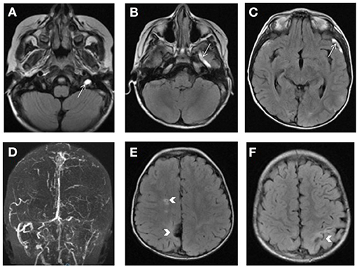
A typical neuroimage findings: (A) In patient 2, hyperintensities ...
Brain MRI after 6 weeks of antifungal treatment. FLAIR (A, D) and T2 ...
Brain MRI findings in children with classical MELAS syndrome. a Brain ...
Isolated Cortical Visual Loss With Subtle Brain MRI Abnormal ...
The cumulative incidences of all CNS complications and PRES at days 30 ...
Bilateral Basal Ganglia Necrosis Secondary to Methamphetamine - Sanchez ...
Posterior reversible encephalopathy syndrome during treatment with ...
T2-weighted MRI images of patient 7, taken at the age of 19 months ...
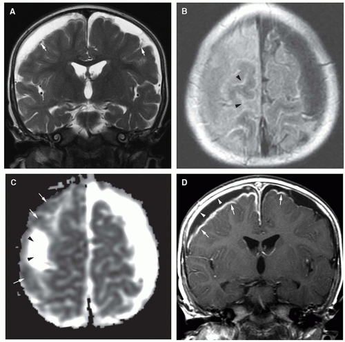
Axial T2-weighted MRI. (A and B) Dot-like hyperintensities ...
Hemichoreo‐hemibalism as a Manifestation of Central Nervous System ...
The severity score of enlarged perivascular spaces in basal ganglia. a ...
Infections of the Developing and Mature Nervous System | Radiology Key
(PDF) Optic Pathway Gliomas in Neurofibromatosis Type 1: An Update
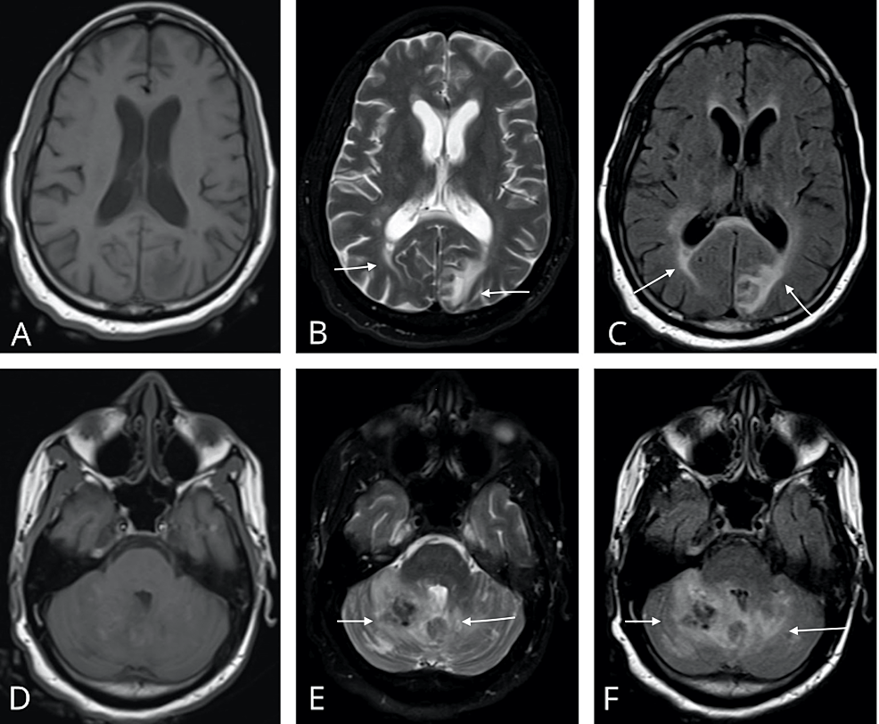
1 MRI of case 1: FLAIR visible hippocampal, thalamic, and midbrain high ...
Cureus | Posterior Reversible Encephalopathy Syndrome With Hemorrhagic ...
Synthetic cannabinoids revealing adrenoleukodystrophy - Journal of ...
PMG. Axial T2-weighted image shows microgyria with normal cortical ...
Teaching NeuroImages: Adrenoleukodystrophy presenting as raised ...
Brain MRI. ( A) Axial T2-weighted image of the brain of patient 1 ...
MRI scans disclosing a well-circumscribed mass in the left frontal ...
Clinical findings of 126 children with familial Mediterranean fever ...
Cerebral Iron Accumulation Is Not a Major Feature of FA2H/SPG35 ...
Representative images of infarcts in multiple cerebrovascular ...
Fast Detection of Diffuse Axonal Damage in Severe Traumatic Brain ...
Parkinsonism & Related Disorders
Hemiballism with leg predominance caused by contralateral subthalamic ...
Cavitation After Acute Symptomatic Lacunar Stroke Depends on Time ...
Evaluation and Treatment of a Patient With Recurrent Stroke in the ...
Case 1. A: T 2-weighted magnetic resonance (MR) image showing an ...
| Hyperintense vessel sign (HVS): (A) Score of 1, restricted to Sylvian ...
A case of chronic progressive lyme encephalitis as a manifestation of ...
Lysosomal Leukodystrophies Lysosomal Storage Diseases Associated With ...
The MRI was performed on the 15th postoperative day, showing the state ...
Ischemic-Hypoxia
Hypoxic-Ischemic-Encephalopathy-MRI
Hypoxic-Ischemic-Brain-Injury
Hypoxic-Ischemic-Encephalopathy-CT
What-Is-Hypoxic-Ischemic-Encephalopathy
Neonatal-Hypoxic-Ischemic-Encephalopathy-MRI
Severe-Hypoxic-Ischemic-Encephalopathy
Hypoxic-Ischemic-Encephalopathy-MRI-Findings
Hypoxic-Ischemic-Encephalopathy-Radiology
Hypoxic-Ischemic-Encephalopathy-Adult
Hie-MRI
Hypoxic-Ischemic-Encephalopathy-Stages
Cerebral-Hypoxia-MRI
Hypoxic-Ischemic-Brain-Damage
Hypoxic-Ischemic-Encephalopathy-in-Newborn
Hypoxic-Ischemic-Encephalopathy-Neonates