Please enter url.
Login
Logout
Please enter url.
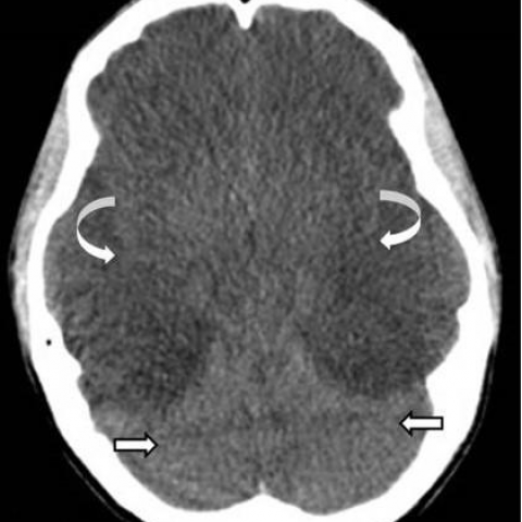
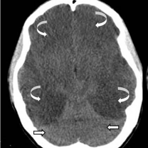
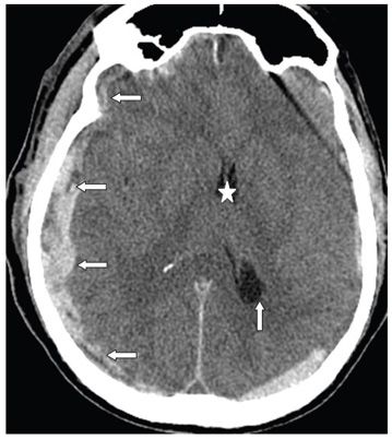
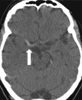
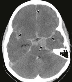
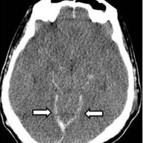
Severe hypoxic - ischaemic brain injury: the reversal, the pseudo ...
eurorad.org
source
Comments
(PDF) Case 14694 Severe hypoxic-ischemic brain injury: the reversal ...
Severe hypoxic - ischaemic brain injury: the reversal, the pseudo ...
Imaging of Vascular and Endovascular Surgery | Radiology Key
(A, B): Hot cross bun sign. Axial T2W MRI image of the pons (A) in a ...
(PDF) Imaging of complicated frontal sinusitis
-Enhanced coronal brain CT shows a growing mass centred in the anterior ...
(PDF) Hyperdense middle and anterior cerebral arteries: Familiar and ...
-CT head showing space occupying lesion involving right frontal region ...
Multidetector-row computed tomography in cerebral hydatid cyst ...
218 | Radiology Key
A 68-year-old woman with a left hemiplegia following a conscious ...
Head Injury and Facial Trauma | Clinical Gate
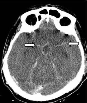
Severe hypoxic - ischaemic brain injury: the reversal, the pseudo ...
287 | Radiology Key
142 | Radiology Key
Osmotic demyelination syndrome (ODS)—extrapontine myelinolysis (EPM ...
-Axial noncontrast CT soft tissue window (a) at the level of skull base ...
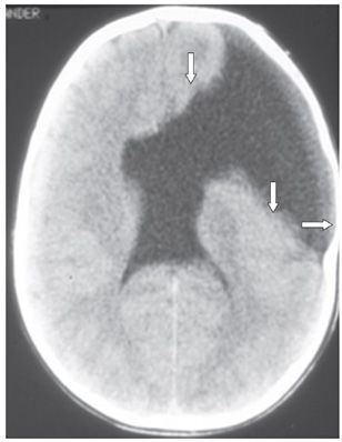
Sturge-Weber syndrome | MedLink Neurology
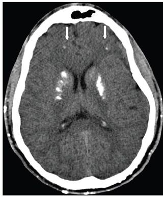
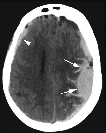
Severe hypoxic - ischaemic brain injury: the reversal, the pseudo ...
167 | Radiology Key
Hypertensive Encephalopathy | AcrossPG Blog!
Non-contrast-enhanced axial CT image showing a hyperdense left anterior ...
Imaging of Cerebrovascular Disease in Pregnancy and the Puerperium | AJR
Common Neuroradiological Procedures | Neupsy Key
The native CT scan of the brain revealed a subtle oval hypodensity on ...
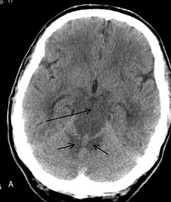
Severe hypoxic - ischaemic brain injury: the reversal, the pseudo ...
Severe hypoxic - ischaemic brain injury: the reversal, the pseudo ...
A 53-year-old man who suffered cardiac arrest secondary to acute ...
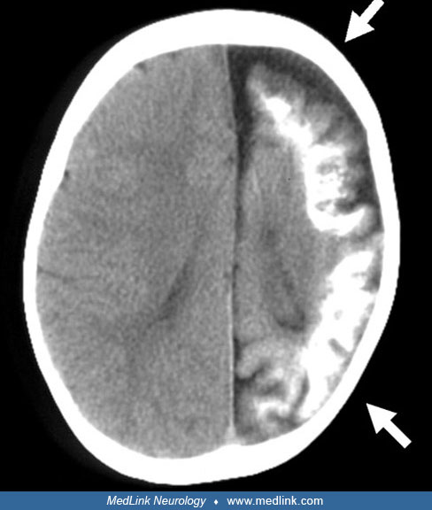
Cerebral oedema and dural sinus thrombosis in an adolescent with ...
Initial CT scan of a 53 year-old man revealing a hyperdense region in ...
42-year-old male with meningitis. MRI of the brain. Note marked ...
RM de cerebro obtenida al séptimo día A) corte axial, FlaiR ...
a. Axial noncontrast head CT image shows a crescent-shaped hyperdense ...
Ependymal Brain Cyst with Posterior Cerebral Artery Infarct | SpringerLink
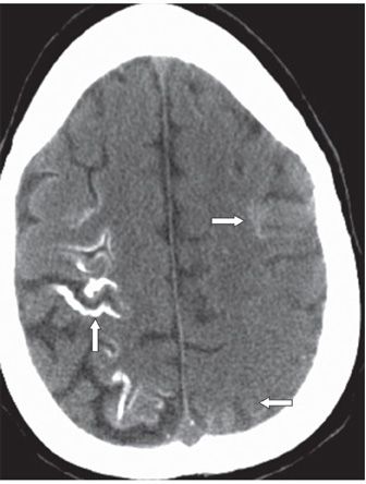
Transverse sinus flow gap. (a) Coronal time-of-flight MR venogram shows ...
CTC-Global
CT-Group-Logo
Global-Technologies
Global-Logistics-Logo
CT-Institute
Windows-Admin-Center-Logo
CT-Global-Freight
Crystal-Mall-Waterford-CT
Citi
Biosimilar-Market
RT-PCR-Report
Global-Resources
A-Logo-PNG
Global-Real-Estate
Global-Atlantic
Connecticut-On-Map