Please enter url.
Login
Logout
Please enter url.
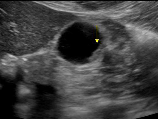
Carotid Siphon Ultrasound
mavink.com
source
Comments
Simplified schematic illustration of the pathophysiologic process of ...
Intralesional blood vessels in gallbladder lesions on CEUS (arrows). A ...
Intrauterine Linear Echogenicities in the Gravid Uterus: What ...
Ocular Ultrasound of a normal eye. The ultrasound probe is applied to ...
Prenatal ultrasonography demonstrated a cystic mass with a thin and ...
TransVaginal USS Image of Caesarean scar pregnancy (CSP). A) Arrow ...
Scheme of the subtotal duodenectomy: the parts of the duodenum to be ...
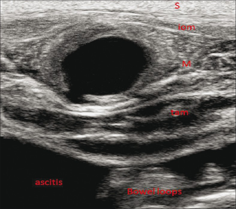
Anterior pelvic abscess seen on ultrasound (a) as complex fluid ...
Esophageal intra mural pseudocyst with wall thickening (arrows ...
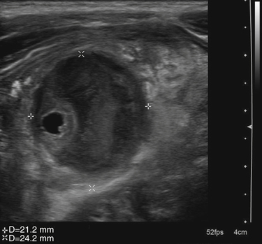
Abdominal ultrasonography demonstrating a left ovary within a canal of ...
Tumefactive sludge. A tumefactive sludge (arrow) presents with polypoid ...
Verification of correct central venous catheter placement in the ...
Colloid nodule. Transverse US image shows a predominantly anechoic ...
(PDF) Dimensional analysis of the endometrial cavity: How many ...
Neonatal Spinal Dimple | Radiology Key
Male Genital Tract | Radiology Key
Abdomen and retroperitoneum | 1.2 Gallbladder and bile ducts : Case 1.2 ...
Deep Circumflex Iliac Artery Pseudoaneurysm as a Complication of ...
Transabdominal ultrasound shows mild focal gallbladder wall thickening ...
Role of POCUS in Acute Appendicitis | Point-of-Care Ultrasound ...
Prenatal Diagnosis of Retinoblastoma - Advances in Ophthalmology and ...
Abdomen and retroperitoneum | 1.2 Gallbladder and bile ducts : Case 1.2 ...
High-frequency transducer gray-scale sonogram of the left testis ...
Normal endoscopic ultrasound image of the pancreas acquired with a ...
Example of the measurement of the optic nerve sheath diameter. Optic ...
(PDF) Endovascular Aneurysm Repair for Abdominal Aortic Aneurysm: A ...
Transrectal Ultrasonography of the Prostate Technique: Transrectal ...
B-scan ultrasonography showing clearly demonstrable calcific densities ...
Gray-scale ultrasound (a) shows well defined, solid mass that causes ...
EPOS™
(PDF) Role of Endoscopic Ultrasound in Gastroenteropancreatic ...
Sonogram of conjoined teat with independent Teat cistern and Gland ...
Epididymal obstruction. Longitudinal image of the right scrotum shows ...
Lens dislocation | Samantha Salesny MD & Maninder Singh MD | Bronx, NY ...
e Sagittal T2-weighted imaging showing a gestational sac (curved white ...
Carotid-Artery-Dissection-Ultrasound
Internal-Carotid-Dissection
Cervical-Carotid-Artery
Carotid-Artery-CT-Scan
Aortic-Intimal-Flap
Common-Carotid-Artery-Dissection
Carotid-Artery-Surgery-Procedure
Intimal-Flap-Aorta
Carotid-Artery-Dissection-CTA
Carotid-Artery-Occlusion
Carotid-Intimal-Thickening
Bilateral-Carotid-Arteries
Carotid-Artery-Plaque-Ultrasound
Right-Carotid-Artery-Dissection
Carotid-Artery-Dissection-MRI
MRA-Carotid-Arteries