Please enter url.
Login
Logout
Please enter url.
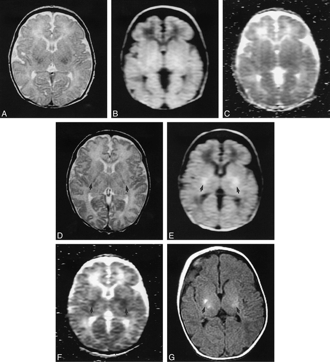
Axial magnetic resonance images in a 29-day-old boy with clinical ...
researchgate.net
source
Comments
MR Line-Scan Diffusion-Weighted Imaging of Term Neonates with Perinatal ...
Axial magnetic resonance images in a 3-month-old girl to evaluate ...
CT and MRI Brain Features of Individuals with Mutations in PROSC (A ...
Assessment of brain tissue injury after moderate hypothermia in ...
Figure 1 from Phenotypic Features of Cerebral Autosomal-Dominant ...
Artifacts secondary to the shunt reservoir. The SS-GRE MR image is the ...
Patient 1: facial features with down-slanting palpebral fissures ...
Clinical and Diagnostic Features of Delayed Hypoxic Leukoencephalopathy ...
Cognitive and Physiologic Correlates of Subclinical Structural Brain ...
Neonatal MRI to Predict Neurodevelopmental Outcomes in Preterm Infants ...
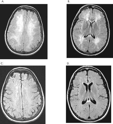
Brain MRI made at the age of 13 months (images A, B, and C) and at the ...
Preeclampsia Eclampsia - Cytotoxic Edema - Mussen Healthcare
Bupropion Overdose Presenting as Status Epilepticus in an Infant ...
Reversible Extralimbic Paraneoplastic Encephalopathies With Large ...
West Nile Virus: Case Report with MR Imaging Findings | American ...
Left peritrigonal focal WM lesion (arrows) appearing hyperintense on ...
The Hyperdense Posterior Cerebral Artery Sign | Stroke
Variable Agreement Between Visual Rating Scales for White Matter ...
Figure 1 from Diagnosing variant Creutzfeldt-Jakob disease with the ...
Diagnosing Variant Creutzfeldt-Jakob Disease with the Pulvinar Sign: MR ...
Diffuse lesion in the splenium of the corpus callosum in patients with ...
Illustrative proton density ( A and C ) and T2 ( B and D ) axial MR ...
The spectrum of associated brain lesions in cerebral sinovenous ...
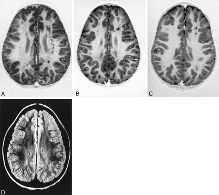
MRI appearance of white matter changes in axial sections of patients ...
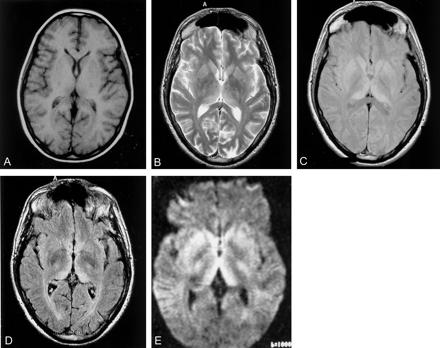
Initial (A, D) and two follow-up magnetic resonance images 3 days (B ...
Blood-brain barrier (BBB) breakdown and neurological complications. (A ...
Chronic Traumatic Encephalopathy | Neupsy Key
Axial FLAIR (A) and diffusionweighted images (B) from the initial MR ...
Case 317 | Radiology
Imaging Inflammation in Acute Brain Ischemia | Stroke
Multiple System Atrophy Ubu - Parkinson Disease - RR School Of Nursing
Figure 1 from Magnetic Resonance Imaging Features in Japanese ...
MRIs of the two epilepsy brothers. A, B, C (case 1) The T2-weighted ...
Axial T2-weighted MRI. (A and B) Dot-like hyperintensities ...
Fig 1. | Cognitive Impairment in Children with Hemoglobin SS Sickle ...
Gray-White-Matter-Differentiation
Grey-White-Matter-Differentiation
Cell-Differentiation-Diagram
Gray-and-White-Matter-Brain
Grey-vs-White-Matter-Brain
Preserved-Gray-White-Differentiation
Loss-of-Gray-White-Matter
Cytotoxic-Edema-Gray-White-Differentiation
Gray-White-Differentiation-MRI
Normal-Gray-White-Differentiation
Gray-Matter-CT
Stem-Cell-Differentiation
Grey-White-Matter-Interface
Grey-White-Matter-Differentiation-Head-CT
Contrast-Gray-and-White-Matter
Grey-White-Matter-Differentiation-in-CT-Scan