Please enter url.
Login
Logout
Please enter url.
Severe Iatrogenic Lutembacher Syndrome in a Young Male: Case Report and ...
medicalresearchjournal.org
source
Comments
Cureus | Advanced Pregnancies With Valvular Heart Disease Requiring ...
Figure 2 from A Rare Presentation with Angina and Pseudoinfarct Ecg ...
Pulmonary stenosis before surgery. | Download Scientific Diagram
A Thread from @ArgaizR: "Pt w ischemic cardiomyopathy. Formal Echo from ...
Figure 1 from Relationship between Perioperative Left Atrial Appendage ...
Transthoracic echocardiography apical four-chamber view showing ...
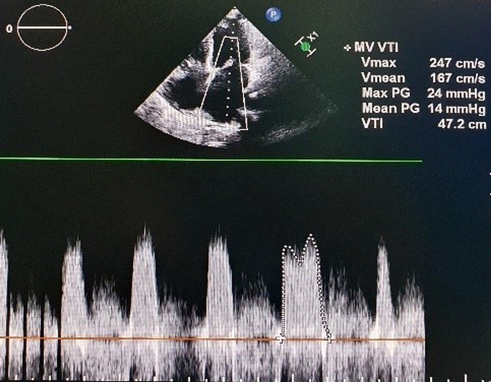
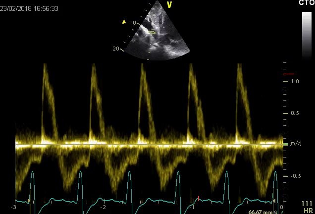
Continuous-wave Doppler with Valsalva maneuver on TTE in the apical ...
Figure 3 from Recurrent Hemopericardium With Cardiac Tamponade as an ...
Left Atrial Appendage, Intraoperative Echocardiography, and the ...
Evaluation of pulmonary arterial stiffness (PAS). PAS = MFS/PAT MFS ...
2012 Philips iE33 Diagnostic Ultrasound System – AusChoice
Two-dimensional midesophageal long axis transesophageal... | Download ...
This image demonstrates staccato flow in the renal artery with absent ...
Kauvery Hospital (Alwarpet) Performs India's First Case of Intracardiac ...
New PHILIPS EPIQ 7C Ultrasound Machine Ultrasound General For Sale ...
The Atria Are Stunned, but You Shouldn't Be
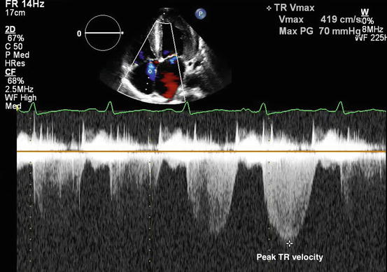
Deep transgastric long-axis view showing a peak gradient of 77 and a ...
Severe Functional Tricuspid Stenosis Secondary to a Giant Saphenous ...
Figure 4. 2 D Echocardiogram with M mode showing spike like projections ...
M mode of echocardiograph showing increased gradient across aortic ...
Ultrasound Course | Online Ultrasound Course | Online Ultrasound ...
Severe AR -with PNT 44 ms | Download Scientific Diagram
(a), (b) Transthoracic echocardiogram (apical (a) and parasternal ...
Cureus | A Rare Case Presentation: Diagnosing Primary Biliary ...
Unilateral Pulmonary Edema Secondary to Mitral Valve Perforation ...
Alessandro Castiglioni on LinkedIn: Minimally invasive mitral valve ...
Figure 10 from Left Ventricular Outflow Tract Obstruction Following ...
Hemodynamics
Echocardiogram 5 weeks post imatinib: (a) 2D parasternal long axis view ...
Assessment of Primary Mitral Valve Disease: Clinical Presentation ...
Echo: Doppler III | Anesthesia Key