Please enter url.
Login
Logout
Please enter url.
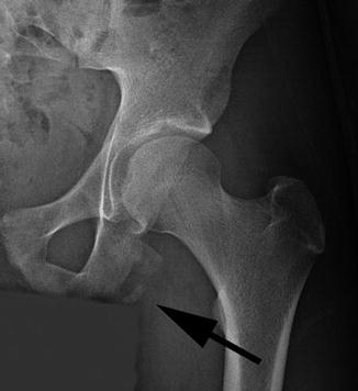
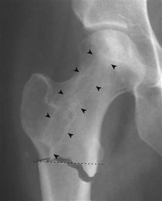
Greater Trochanter Avulsion Fracture
mungfali.com
source
Comments
MRI scan showing an avulsion of the right lesser trochanter. | Download ...
Supine AP X-ray of the right hip joint with a non-displaced subcapital ...
A frontal radiograph of the left hip featuring a coxa profunda which is ...
Radiografia pós-operatória de 2 anos em incidência anteroposterior de ...
Hand and wrist radiographs (d) show typical rachitic changes in ...
a-Acetabular dysplasia of the right hip in a 15-year-old girl ...
Evaluation of the Child with a Limp | 2004-03-01 | AHC Media:…
Cureus | Osteoid Osteoma at the Lesser Trochanter: A Lesson in Mimicry
The initial MRI scan of the patient’s pelvis. Short TI Inversion ...
(A) Anteroposterior pelvis radiograph demonstrating an os acetabuli ...
Pelvic AP radiograph showing PRIS 1 and PRIS 2 measurements. | Download ...
Femoral Head Sparing Procedures for Osteonecrosis of the Hip ...
Acetabular Lipping | Sitelip.org
Pelvic and hip abnormalities associated with DMC syndrome. The pelvis ...
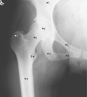
Hip X-ray Landmarks Diagram | Quizlet
[PDF] Acute severe hip pain associated with labral calcific deposition ...
Slipped upper femoral epiphysis | Eurorad
(A) An anterior posterior pelvic radiograph of a 21-year-old patient ...
Hip pain in a middle aged woman | The BMJ
Femur Radiograph #2 Diagram | Quizlet
Absolute Risk for Femur Fracture Low With Bisphosphonates
Femoroacetabular Impingement in Athletes | Musculoskeletal Key
Anterior-posterior radiograph of the pelvis showing patchy ...
frog lateral hip Diagram | Quizlet
Limb | Basicmedical Key
a/p X-ray one week after closed reduction shows a congruent hip joint ...
Image | Radiopaedia.org
Art. coxae Diagram | Quizlet
A 15-year-old female patient with hip pain
Surgical Technique: Endoscopic Hamstring Repair and Ischial Bursectomy ...
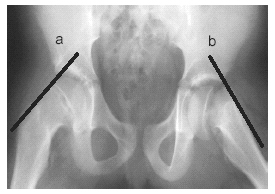
Judet Views - wikiRadiography
:: JKSR :: Journal of the Korean Society of Radiology
A frontal radiograph of the left hip featuring a coxa profunda which is ...
Imaging in Osteomalacia and Renal Osteodystrophy: Practice Essentials ...
CT scan shows right hip dislocation and dysplastic acetabulum and there ...
Avulsion-Fracture-Tibia
Tibial-Avulsion-Fracture
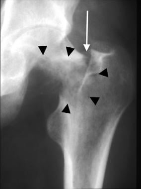
Lesser-Trochanter-Avulsion
Ischial-Avulsion-Fracture
Lesser-Trochanteric
Trochanteric-Hip-Fracture
Apophyseal-Avulsion-Fracture
Ischial-Tuberosity-Avulsion-Fracture
Pelvic-Avulsion-Fracture
Aiis-Pelvis
Tibial-Tubercle-Avulsion-Fracture
Avulsion-Fracture-Shoulder
Greater-Trochanter-Hip-Fracture
Intertrochanteric-Hip-Fracture
Rectus-Femoris-Avulsion-Fracture
Hip-Flexor-Avulsion-Fracture


![[PDF] Acute severe hip pain associated with labral calcific deposition ...](https://d3i71xaburhd42.cloudfront.net/d385f285fe4ef8ab91c62763f9a87e9cb5d9487e/250px/2-Figure1-1.png)