Please enter url.
Login
Logout
Please enter url.
Cureus | Prosthetic Mitral Valve Obstruction with Left Atrial Appendage ...
cureus.com
source
Comments
Cureus | Prosthetic Mitral Valve Obstruction with Left Atrial Appendage ...
Sub-aortic at the admission, first patient | Download Scientific Diagram
(a) Abnormal pulsatile flow with a retrograde component in saphenous ...
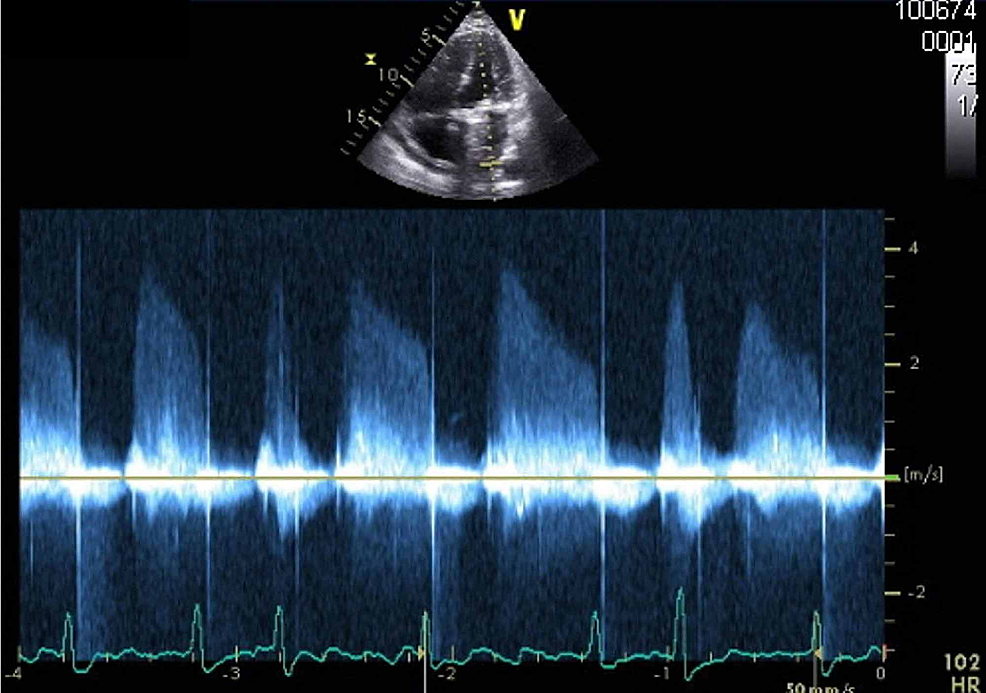
Transmitral inflow traces showing signicicant respiratory phase ...
Apical short-axis view, TTE: showing a mitral valve E/A ratio of 0.75 ...
Right ventricular myocardial performance index (RVmpi') assessed by ...
Pulmonary artery systolic pressure | Download Scientific Diagram
(PDF) Echocardiographic Evaluation of Tricuspid Prosthetic Valves: An ...
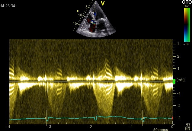
Typical appearance of a PSW on pulsewave Doppler examination of the ...
Transthoracic echocardiography showing continuous‐wave Doppler at the ...
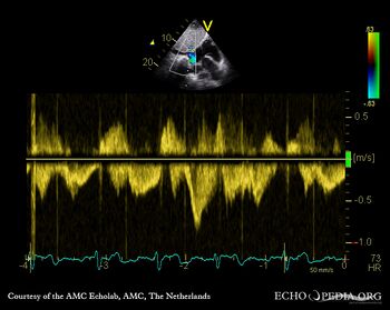
Case 97 - Echopedia
Transthoracic left apical window modified 4-chamber view optimized for ...
Pulmonary pulse transit time is defined as the time interval between ...
Lunate Dislocation - JETem
Figure 1 from Respiratory variation in aortic flow peak velocity and ...
Apical 4-chamber view (right heart centered) during stress ...
Transthoracic left apical window modified 4-chamber view optimized for ...
Normal mitral inflow pattern. E>A, E-early inflow wave: A-atrial ...
Pulsed Doppler sample volume was placed at the mitral valve tip, and ...
Ultrasound Animal Challenge 1 – What is the Tiger-stripe sign ...
Supraventricular trigeminy (arrows show ectopic atrial beats ...
Medial early diastolic mitral annular velocity (e′) measured by ...
Determining fetal heart rate using pulsatile Doppler [positive wave ...
Ventricular Septal Defect (VSD) - Echopedia
Systolic anterior motion (SAM) of the mitral valve visualized by M-Mode ...
Levosimendan for management of cardiogenic shock with electrical storm ...
Representative ECG and PCG signals showing the PEP and LVET time ...
(PDF) Trilogy of Fallot: a rare congenital heart disease
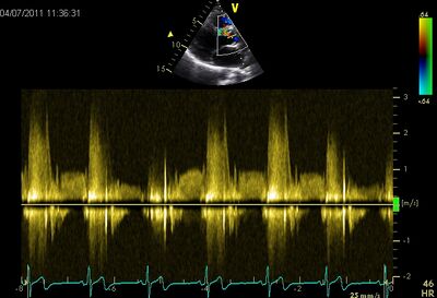
Two-dimensional basal parasternal short-axis view, pulsed-wave Doppler ...
Acquired mitral valve aneurysm due to severe aortic regurgitation ...
-Continuous wave Doppler of the aortic valve showed severe aortic ...
Transthoracic echocardiogram-continuous wave Doppler demonstrating ...
A Pericardial thickening in the long axis of the apex, anterior to the ...
Transabdominal ultrasound demonstrating of regurgitation across the ...
Preoperative measurement of gradient of aortic regurgitation and AS ...
Four-Chamber-View-Echo
Subcostal-4-Chamber-View
2-Chamber-View
Apical-4-Chamber-Echo
Apical-3-Chamber-View
5-Chamber-View-Echo
Pulmonary-Veins-Echo
Transthoracic-Echo-Views
Normal-4-Chamber-Echo
4-Chamber-Heart-Ultrasound
4-Chamber-Heart-Diagram
Fetal-4-Chamber-Heart-Ultrasound
Apical-4-Chamber-Wall-Segments
Tee-4-Chamber-View
Perimembranous-VSD-Echo
Fetal-Outflow-Tracts-Ultrasound