Please enter url.
Login
Logout
Please enter url.
Ulnar Collateral Ligament Wrist Mri
animalia-life.club
source
Comments
Sensors | Free Full-Text | Motion Analysis of Triangular Fibrocartilage ...
25-year-old woman with wrist pain - Auntminnie
Imaging of the wrist at 1.5 tesla using isotropic three‐dimensional ...
Radiographic findings of ulnar impaction syndrome in fishery workers ...
Silicone radial head prostheses: the clinical course and treatment of ...
Figure 1 from Remodelling of the distal radius after epiphysiolysis and ...
Imaging of physeal bars in children | SpringerLink
What is an MRI wrist arthrogram? - YouTube
Application of 3 dimension-printed injection-molded polyether ether ...
JCM | Free Full-Text | Pathomechanism of Triangular Fibrocartilage ...
Arthroscopic one-tunnel transosseous foveal repair for triangular ...
Image | Radiopaedia.org
Cancers | Free Full-Text | State of the Art and New Concepts in Giant ...
Preiser’s Disease—Current Concepts of Etiology and Management - Hand ...
Distal Radius and Carpal Fractures | Obgyn Key
CHRONIC DRUJ problem. A chronic DRUJ problem (stabilized then ...
A Scapholunate Ligament–Sparing Technique Utilizing the Medial Femoral ...
Figure 2 from Dorsal Scaphoid Subluxation on Sagittal Magnetic ...
Microorganisms | Free Full-Text | Pediatric Osteoarticular Kingella ...
Cancers | Free Full-Text | State of the Art and New Concepts in Giant ...
Trapeziectomy | Radiology Case | Radiopaedia.org
Kienbock disease | Image | Radiopaedia.org
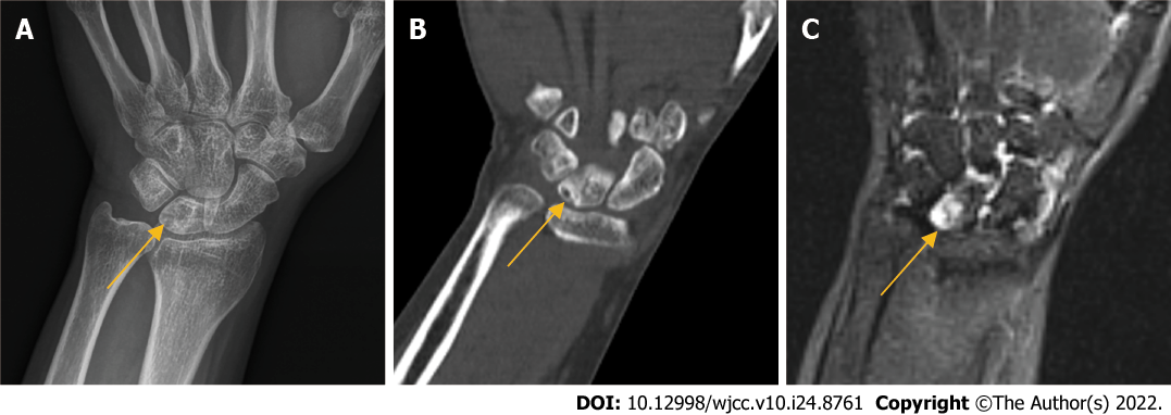
Plain X-ray of the left carpus corresponding to case 3 which shows ...
Die-punch fracture | Image | Radiopaedia.org
Repair of Arthroscopic Triangular Fibrocartilage Complex Tears in ...
Coronal PD FS-MAGNETOM Free.Max - Siemens Healthineers USA
Image | Radiopaedia.org
Free-Vascularized Medial Femoral Condyle Bone Transfer in th ...
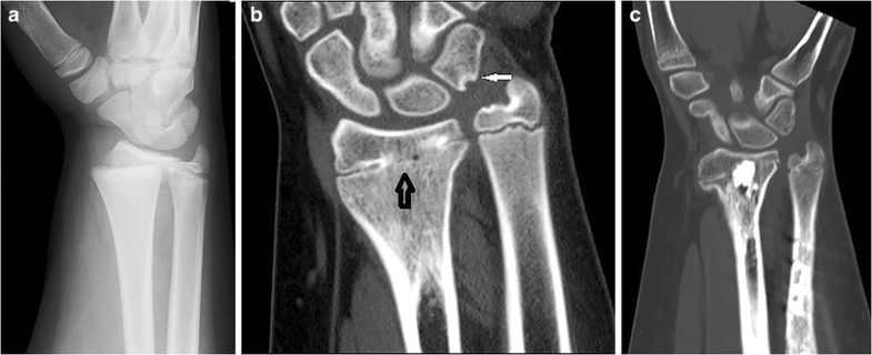
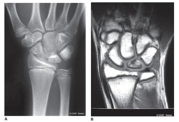
ap and lateral plain radiographs and coronal CT of initial imaging of ...
Image | Radiopaedia.org
(a,b,c,d) Radiographs and computerised tomography 3 months after ...
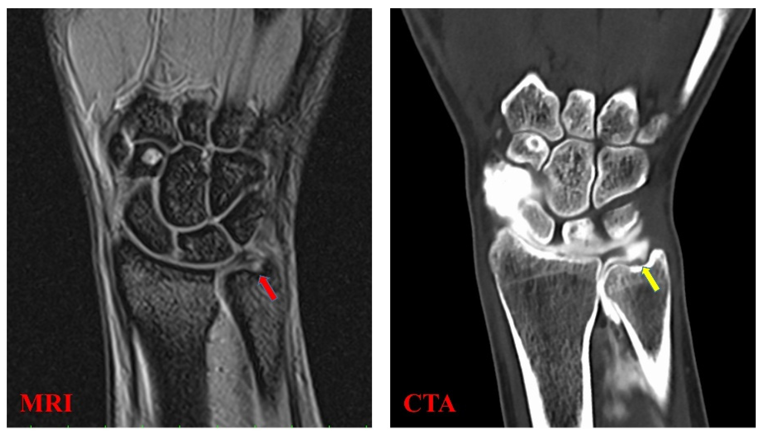
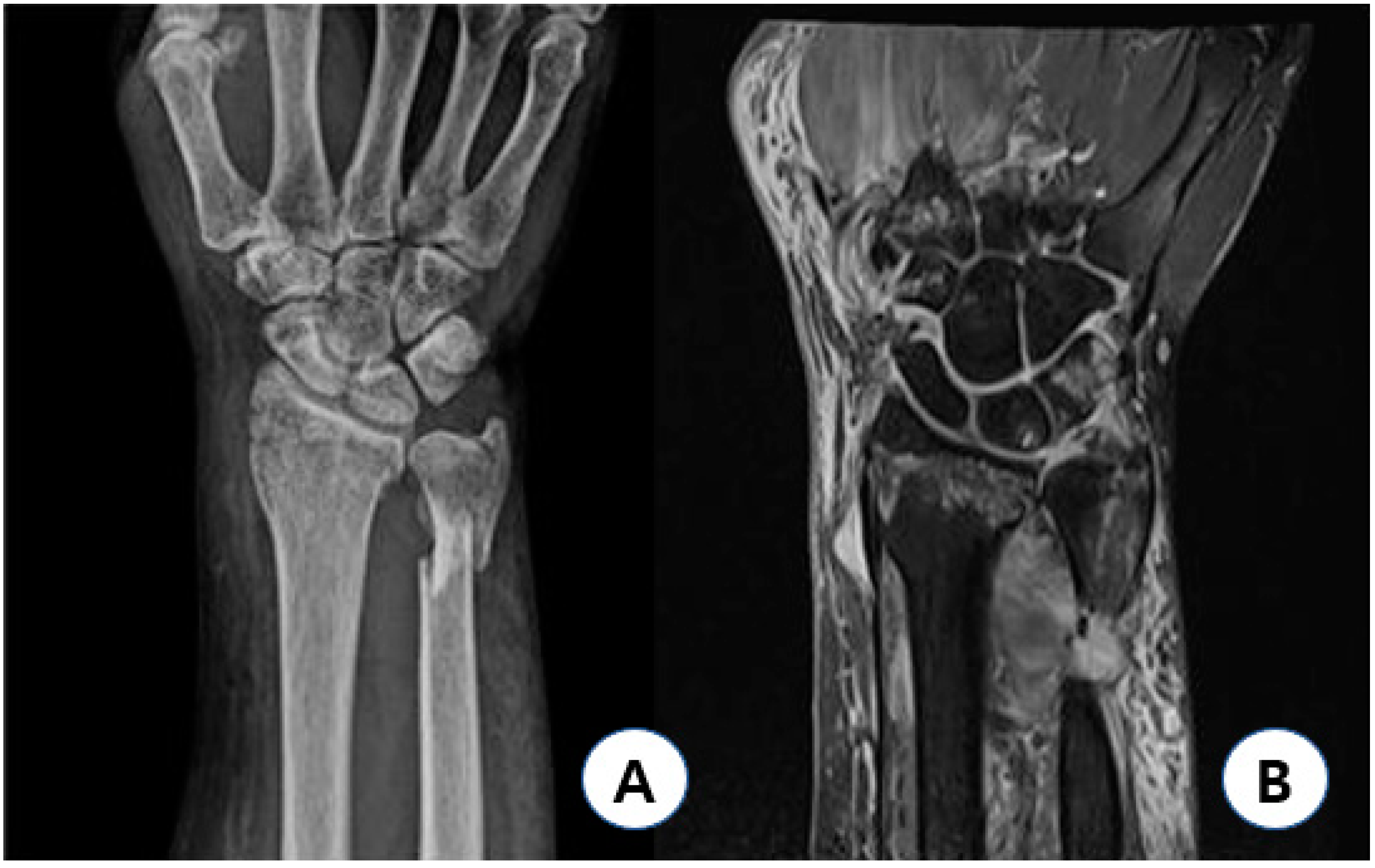
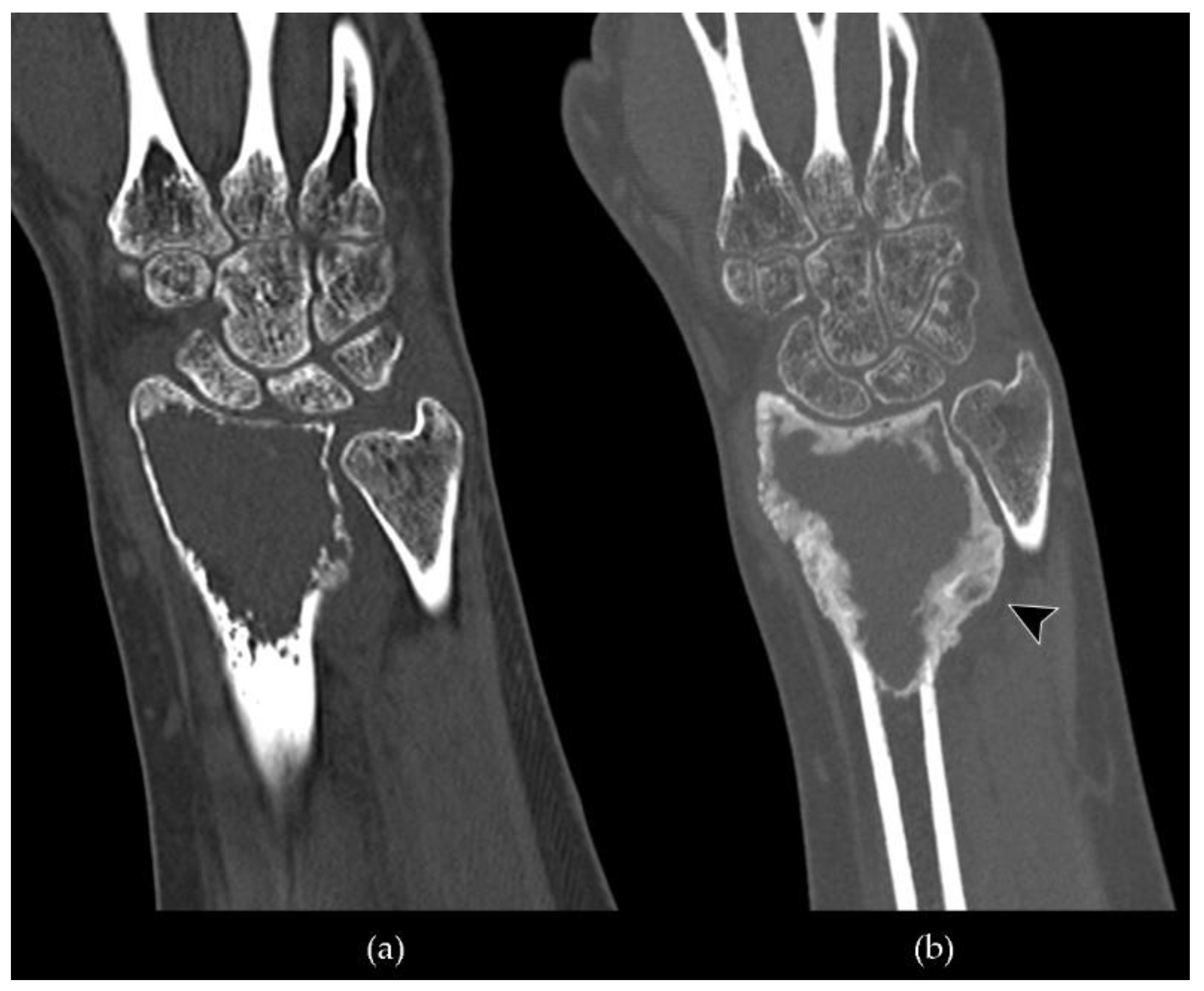
A Representative view of the right wrist on CT scan. B Representative ...
Distal Radioulnar Joint: Normal Anatomy, Imaging of Common Disorders ...