Please enter url.
Login
Logout
Please enter url.
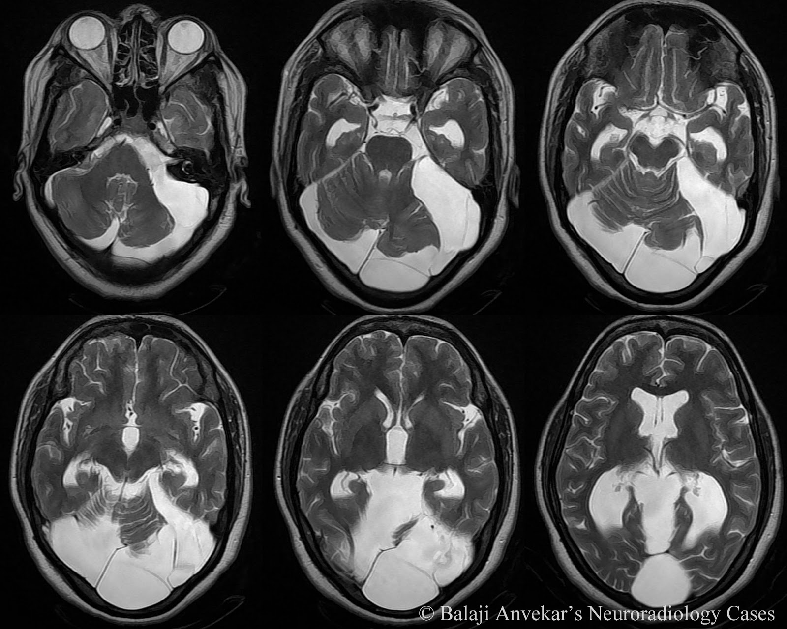
T2 and FLAIR hyperintensity in a patient with Machado-Joseph disease ...
researchgate.net
source
Comments
The multiple faces of encephalitis: Antibody profile in a case series ...
Axial T2-weighted magnetic resonance images revealing multifocal ...
Wilson's Disease: Clinical Practice Guidelines of the Indian National ...
Oculodentodigital Dysplasia: A Hypomyelinating Leukodystrophy with a ...
Challenges in Diagnosing Comatose Patients with Ethylene Glycol ...
Brain MRI at 10 months after taking tacrolimus shows a low signal ...
Magnetic resonance imaging enhancement of brain. A-D showed a lesion in ...
Dr Balaji Anvekar FRCR: Posterior Fossa Arachnoid cyst MRI
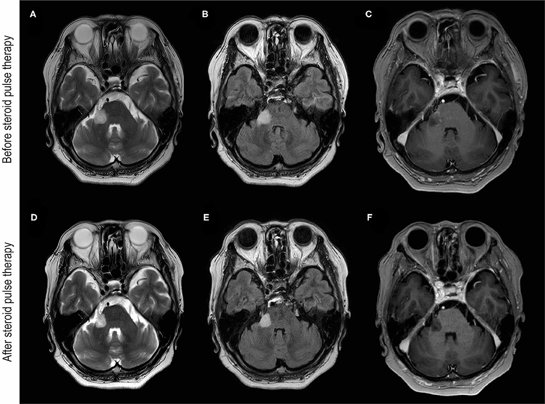
Frontiers | MOG Antibody-Associated Disorders Following SARS-CoV-2 ...
‘Killer’ open ring in the brain | BMJ Case Reports
Brain MRI of P5 aged 23 years. a–e Axial T2-weighted images (axial ...
Frontiers | Case report: Hypnic headache responds to agomelatine–a ...
A novel TRMT5 mutation causes a complex inherited neuropathy syndrome ...
Progressive multifocal leukoencephalopathy, more than an encephalopathy
Mild encephalitis/encephalopathy with reversible splenial lesion (MERS ...
Atypical PLA2G6‐Associated Neurodegeneration: Social Communication ...
Novel compound heterozygous mutations of POLR3A revealed by whole-exome ...
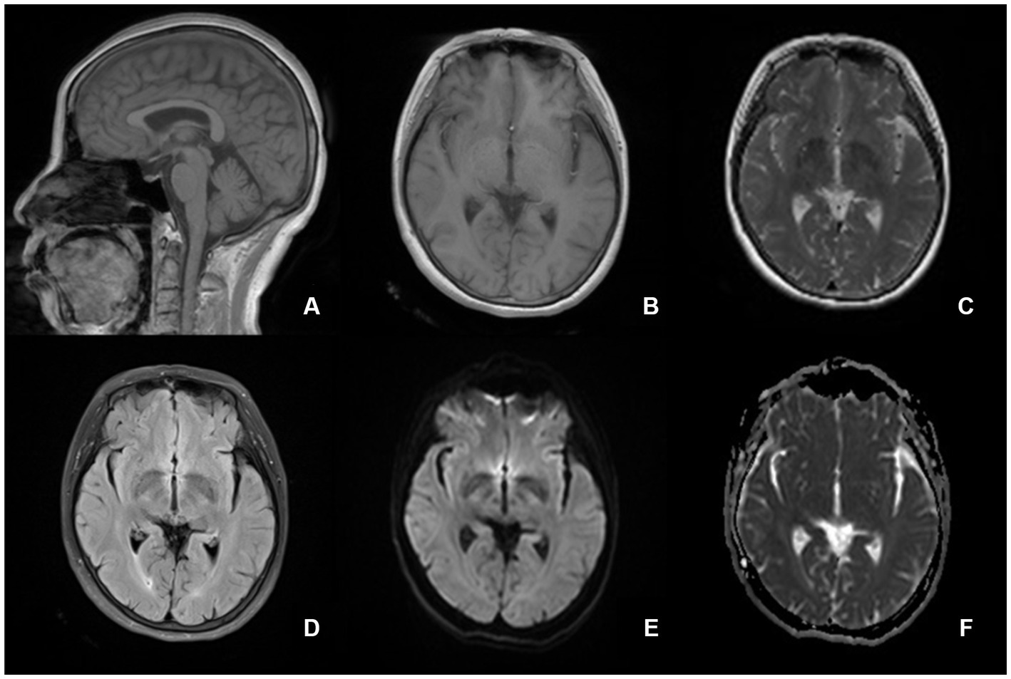
Clinical characteristics of autoimmune GFAP astrocytopathy - Journal of ...
Mild Zellweger syndrome due to functionally confirmed novel PEX1 ...
Severe Early‐Onset Parkinsonian Syndrome Caused by PLA2G6 Heterozygous ...
MRI showed diffuse infiltrative high intensity signal on FLAIR images ...
Clinical Reasoning: A Middle-aged Man With a History of Muscle Pain ...
MRI characteristics of individual 1 Brain MRI of individual 1 at 17 ...
Imaging features of relapsing demyelinating syndromes. | Download ...
A MRI and FLAIR T2W images. Cortical and subcortical high T2 signal in ...
| MRI findings in this case of posterior reversible encephalopathy ...
Aceruloplasminemia and neuroferritinopathy. (a-d) A 60-year-old female ...
Figure 1 from Bilateral posterior cerebral artery stroke following ...
Preoperative MRI. a Coronal T2. b Axial T2. c Axial T2-FLAIR: signal ...
The Mammillary Bodies: A Review of Causes of Injury in Infants and ...
-Brain MRI demonstrates identical signal of the substance in the left ...
RFC1 and FGF14 Repeat Expansions in Serbian Patients with Cerebellar ...
Chiari type I malformation caused by craniometaphyseal dysplasia ...
Figure 1 from A case of multiple sclerosis presenting with inflammatory ...
Posterior reversible encephalopathy syndrome (PRES): diagnosis and ...