Please enter url.
Login
Logout
Please enter url.
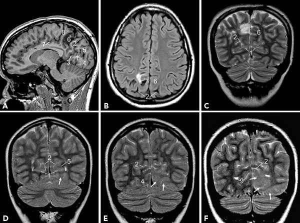
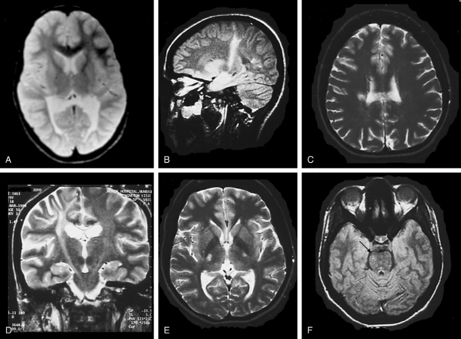
Brain MRI imaging. T2-weighted TSE axial scans (1.5 T) at the level of ...
researchgate.net
source
Comments
Brain MRI imaging. T2-weighted TSE axial scans (1.5 T) at the level of ...
Effects of hematopoietic stem cell transplantation on acyl-CoA oxidase ...
Visual and neurologic sequelae of methanol poisoning in Saudi Arabia ...
MRI findings of crossed cerebellar diaschisis in a case of Rasmussen’s ...
A 47-Year-Old Man Presenting With Seizures and Prior Stroke - Kiara ...
Thalamic deep brain stimulation for acquired dystonia in children and ...
CASE 2: Postnatal brain imaging of the neonates. Postnatal day 7 MRI ...
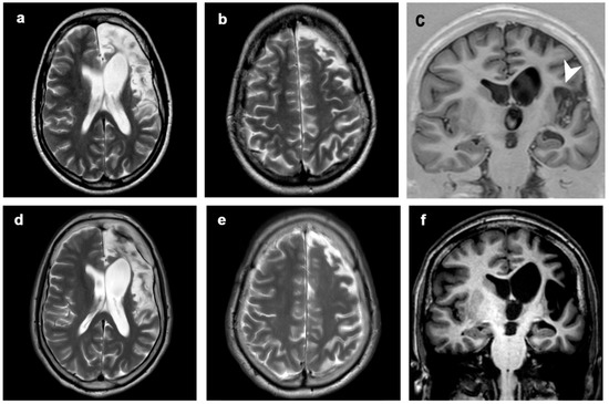
Brain magnetic resonance imaging of patient 1 ( a , b ) and patient 2 ...
Diagnostics | Free Full-Text | MRI in Late-Onset Rasmussen Encephalitis ...
Parkinson's Brain Mri Vs Normal - ParkinsonsInfoClub.com
MEG in mesial ETLE. (A) MEG results from an 18-year-old man with ...
Cerebrale Hilus - Magnetic Resonance - 78 Steps Health Journal
Figure 1 from Moyamoya disease with occlusion of bilateral vertebral ...
Multiple cerebral lesions in a patient with refractory celiac disease ...
Frontiers | Neuroimaging findings in preclinical amyotrophic lateral ...
Prolonged High-Dose Isoflurane for Refractory Status Epilept ...
THE LEUKODYSTROPHIES | Neupsy Key
Figure 2 from A case report of an immunocompetent patient with ...
[PDF] Posterior Reversible Encephalopathy Syndrome With Hemorrhagic ...
MR Imaging Presentation of Intracranial Disease Associated with ...
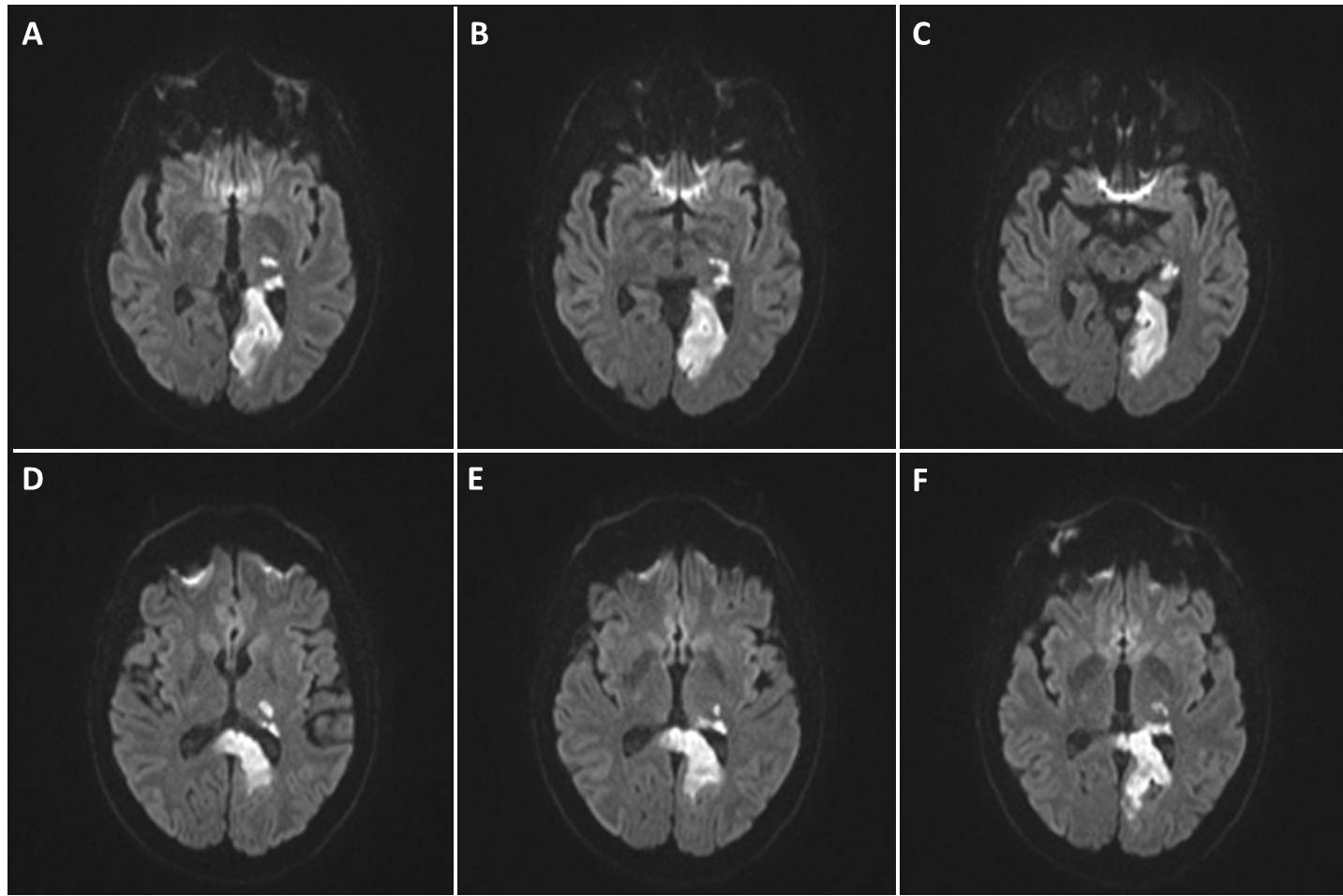
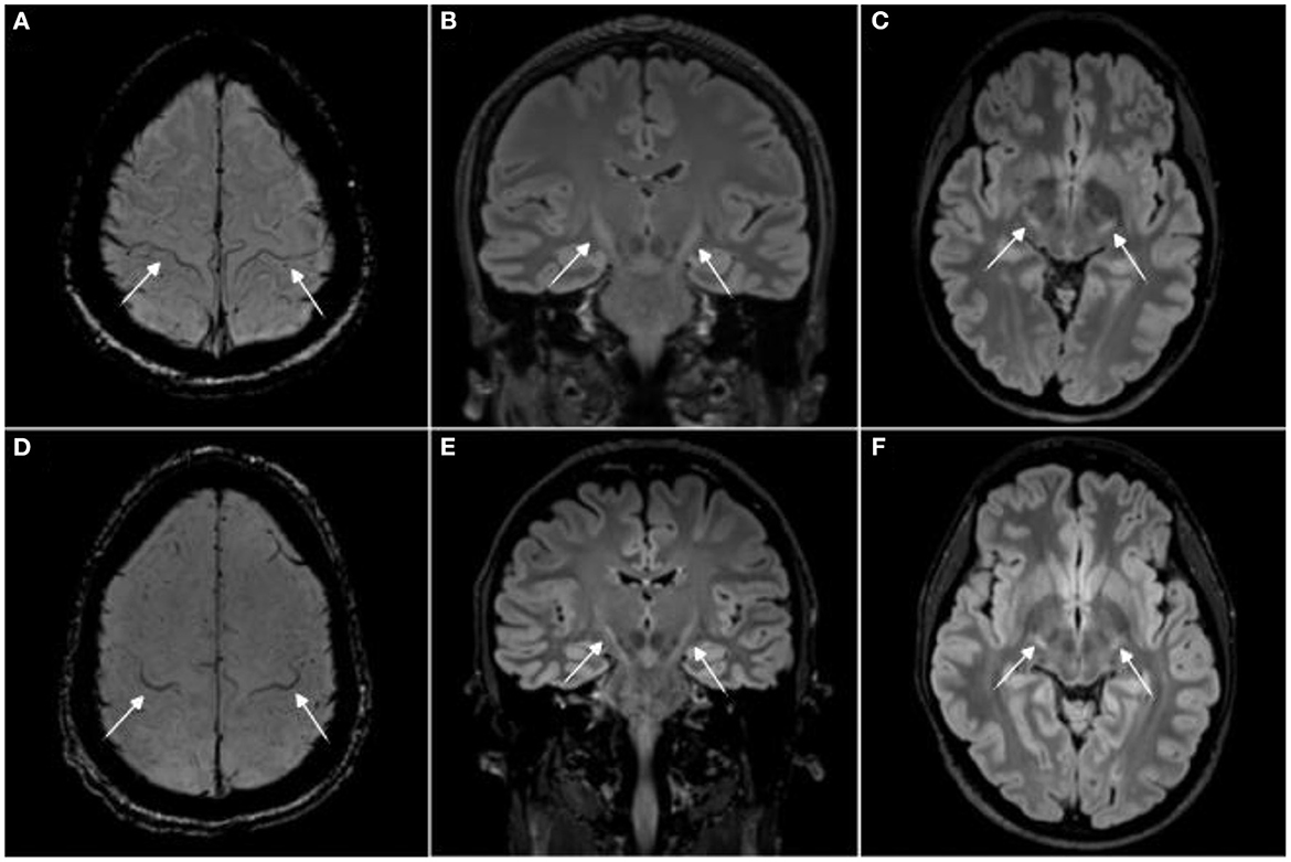
Axial T2 (A–C) and FLAIR (D–F) showing swelling and abnormal high ...
Hemichoreo‐hemibalism as a Manifestation of Central Nervous System ...
Case 4 – preoperative axial T2-weighted MR of the brain demonstrating a ...
Rim enhancing lesion in the left occipital lobe with extensive ...
Magnetic Resonance Imaging in Evaluation for Epilepsy Surgery | Neupsy Key
Magnetic resonance imaging of various categories of tuberculous mass ...
Multiple Myeloma Invasion of the Central Nervous System | Allergy and ...
(A,B) Axial nonenhanced T1-weighted MRI in patient 3, which shows ...
The Mammillary Bodies: A Review of Causes of Injury in Infants and ...
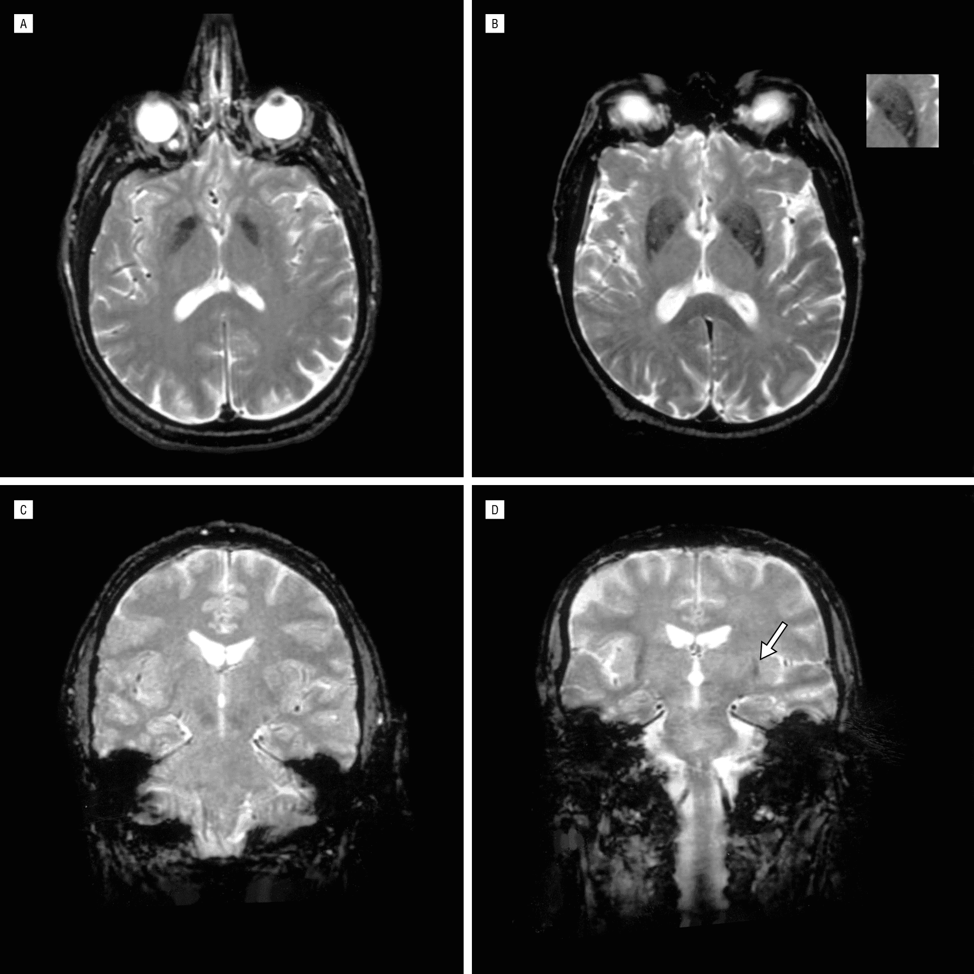
| (a-C) T2-weighted axial magnetic resonance imaging (MRI) revealed ...
Susceptibility weighted imaging in the diagnostic evaluation of ...
Secondary causes of parkinsonism. This figure shows examples of ...
Asymmetric distributed multiple cysts in basal ganglia and corona ...
Preoperative magnetic resonance imaging showing a 1.3 Â 1.2 Â 1.0 cm ...
Image | Radiopaedia.org


![[PDF] Posterior Reversible Encephalopathy Syndrome With Hemorrhagic ...](https://d3i71xaburhd42.cloudfront.net/42553b791acc17880d138fae3a36691f1cd77af4/2-Figure1-1.png)