


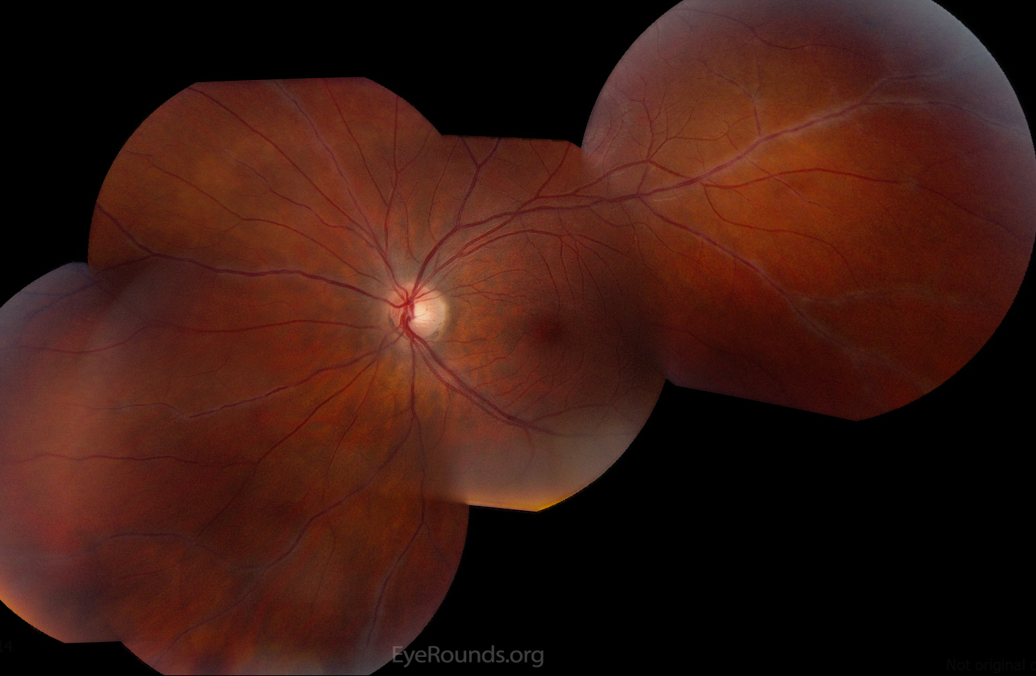


![[Figure, Figure 2: Fundus photography of...] - StatPearls - NCBI Bookshelf](https://www.ncbi.nlm.nih.gov/sites/books/NBK580538/bin/perivasculitis__x1x.jpg)


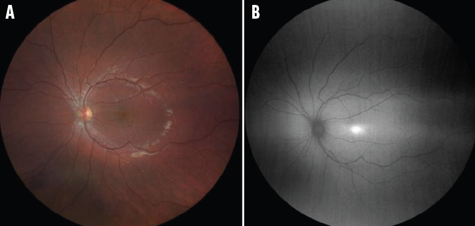


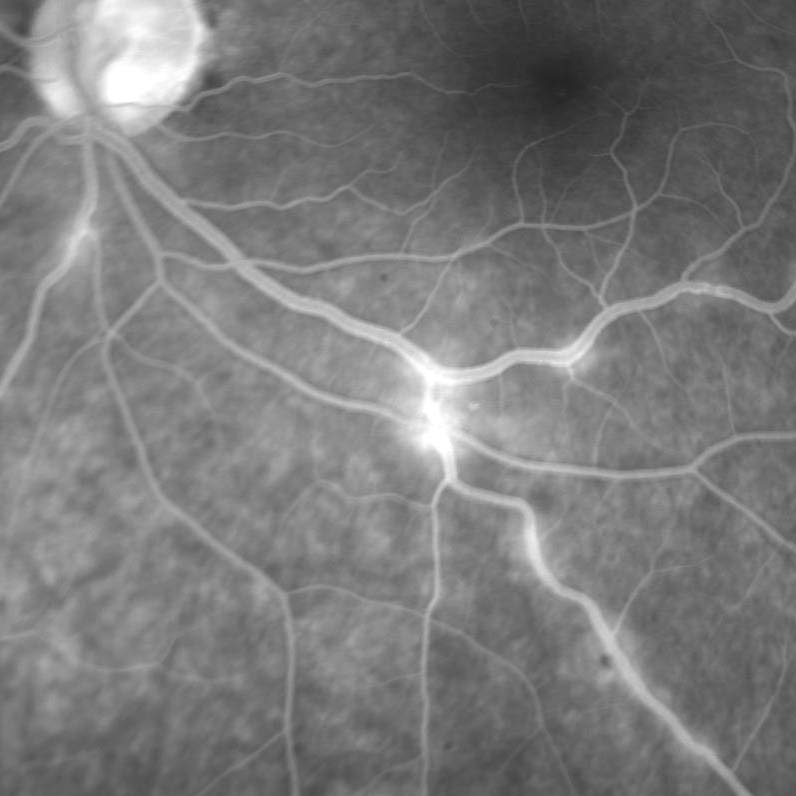
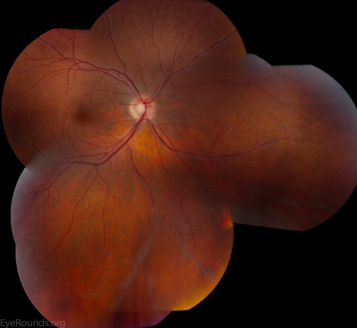
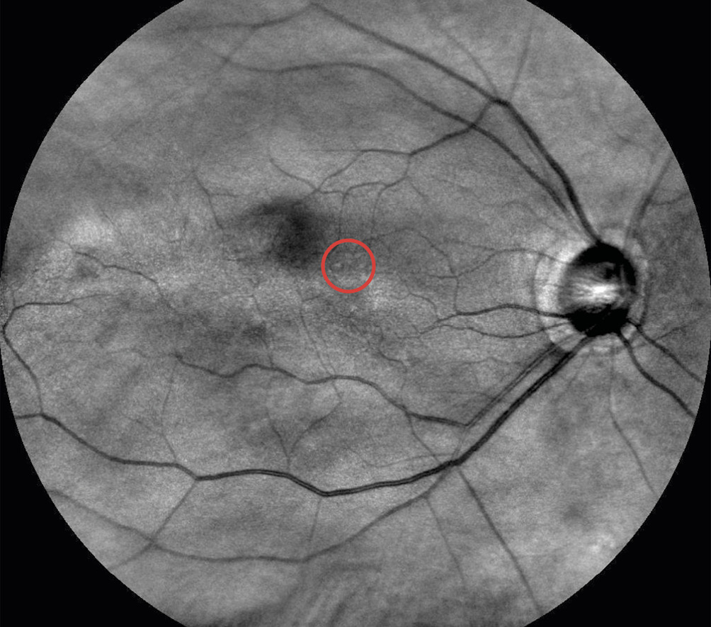
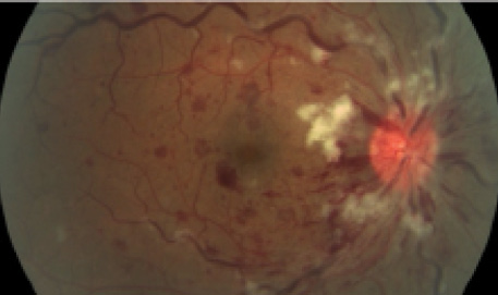
Access our comprehensive in the right eye, fundus imaging (a1) shows peripheral periphlebitis database featuring extensive collections of professionally captured photographs. optimized for both digital and print applications across multiple platforms. providing reliable visual resources for business and academic use. Discover high-resolution in the right eye, fundus imaging (a1) shows peripheral periphlebitis images optimized for various applications. Perfect for marketing materials, corporate presentations, advertising campaigns, and professional publications All in the right eye, fundus imaging (a1) shows peripheral periphlebitis images are available in high resolution with professional-grade quality, optimized for both digital and print applications, and include comprehensive metadata for easy organization and usage. Each in the right eye, fundus imaging (a1) shows peripheral periphlebitis image meets rigorous quality standards for commercial applications. Cost-effective licensing makes professional in the right eye, fundus imaging (a1) shows peripheral periphlebitis photography accessible to all budgets. Instant download capabilities enable immediate access to chosen in the right eye, fundus imaging (a1) shows peripheral periphlebitis images. Our in the right eye, fundus imaging (a1) shows peripheral periphlebitis database continuously expands with fresh, relevant content from skilled photographers. The in the right eye, fundus imaging (a1) shows peripheral periphlebitis collection represents years of careful curation and professional standards.