Please enter url.
Login
Logout
Please enter url.
Parkinson S Disease Brain Region Brain Mri Scans Of A Patient With | My ...
myxxgirl.com
source
Comments
Pin on Fight for Parkinson's
Interpretation Scheme for Nonexpert Pediatricians Evaluating Magnetic ...
The Neurocritic: "Sleeping Beauty Paraphilia" and Body Image ...
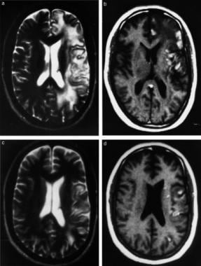
Figure 2 from Serial magnetic resonance imaging changes in hypoglycemic ...
Regional Ischemia and Ischemic Injury in Patients With Acute Middle ...
(abstract A14). A Comparison Across Different Stroke Lesion Profiles at ...
Progression of cerebral white matter lesions: 6-year results of the ...
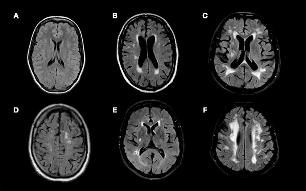
MRI data set in axial views from patient OC are presented (A to F ...
Hereditary diffuse leukoencephalopathy with axonal spheroids caused by ...
RARE MANIFESTATION OF RED INFECTATION: PROGRESSIVE RED PANENSEPHALITE ...
Identification of Major Ischemic Change | Stroke
Unusual Brain MRI Pattern in 2 Patients with COVID-19 Acute Respiratory ...
Cocaine-induced cerebrovascular disease. Magnetic resonance image (MRI ...
Brain Lesions Mri
Primary Angiitis of the CNS Workup: Laboratory Studies, Imaging Studies ...
A and B. Cranial MRI, diffusion sequences showed two right cortical ...
Fluid attenuated inversion recovery (FLAIR) MRI sequences of (A) a 21 ...
Frontiers | Central Nervous System Complications in Children Receiving ...
(PDF) Acute encephalopathy in children with Dravet syndrome
Images of a 54-year-old man with infarction of the left middle cerebral ...
MRI appearance of unilateral right-sided polymicrogyria (PMG ...
Diagnostic criteria for ADEM | Download Table
Frontiers | Fasting Versus Post-Challenge Triglycerides and Pre ...
Fluid attenuated inversion recovery (FLAIR) MRI sequences of (A) a 21 ...
Classification of the Idiopathic Inflammatory Demyelinating Disease of ...
Evolution of common types of preterm brain injury, at 30 weeks ...
(PDF) Investigations of Huntington’s Disease and Huntington’s Disease ...
Data/Images - Adrenoleukodystrophy - ALD
The T2-weighted image (A-C) and apparent diffusion coefficient (ADC ...
Twenty one-year-old man with lymphomatosis cerebri. a, b and c, Axial ...
Figure 1 from Bruxism Secondary to Hypoxic Brain Injury Treated With ...
Case 5. A: T 1 -weighted magnetic resonance (MR) image showing the ...
Angiotensinogen Polymorphism M235T, Carotid Atherosclerosis, and Small ...