Please enter url.
Login
Logout
Please enter url.
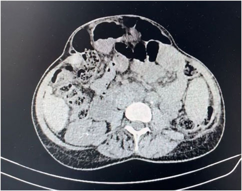
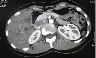
Abdominal CT Scan Lymph Nodes
mungfali.com
source
Comments
Axial CT scan obtained with IV contrast material in 17-year-old male ...
Abdominal cocoon | Eurorad
Contrast injected into the right renal pelvis traveled to the duodenum ...
207 best images about CT Scans on Pinterest | Cars, Gallstone and ...
Castleman’s disease: Mesenteric mass | Eurorad
Pseudomembranous colitis | Radiology Case | Radiopaedia.org
Pin on CT Scans
Acute Epiploic Appendagitis: A Rare Case Report
Abdominal CT scan image showing gallbladder and sigmoid colon process ...
Frontiers Publishing Partnerships | Case Report: Abdominal Wall ...
VIETNAMESE MEDIC ULTRASOUND: CASE 172: HEPATIC ECTOPIC PREGNANCY, Dr ...
Pin by Tnfri on ct bung | Pet ct, Diagnostic imaging, Protected health ...
An abdomen CT scan shows the skin line (the green arrow) and the curved ...
Pin on CT Scans
View of the surgical specimen with the ileal polyp. | Download ...
Sonographic and CT imaging findings of epiploic appendagitis and its ...
-Abdominal CT scan displaying a large left-sided perinephric hematoma ...
A gallstone in the Jejunum and an upstream distension was seen on CT ...
Pin on CT Scans
Abdominal cocoon | Eurorad
Plain computed tomographic scan shows a renal fistula penetrating the ...
Abdominopelvic Anatomy Fig 17-9 Diagram | Quizlet
Cureus | Multidisciplinary Management of Subclavian Artery Perforation ...
Pin on CT Scans
CT Abdomen revealing air-fluid levels, with dilation of multiple loops ...
FULL TEXT - A case of hepatic portal venous gas: When time is gold ...
MRCP showing a dilated and irregular pancreatic duct with intra ductal ...
Salah Ben Elhend, Redouane Roukhsi
CT scan of lumbar hernia (axial view) (blue arrows). | Download ...
(PDF) Stomal stenosis during gradual closure of a traumatic abdominal ...
(PDF) Giant Pelvic Abscess with Sepsis: Case Report and Review of ...
Southwest Journal of Pulmonary, Critical Care and Sleep - Imaging ...
#Interventional #radiologists can use #CT to localize internal # ...
Axial CT scan demonstrating a distended appendix with surrounding fat ...
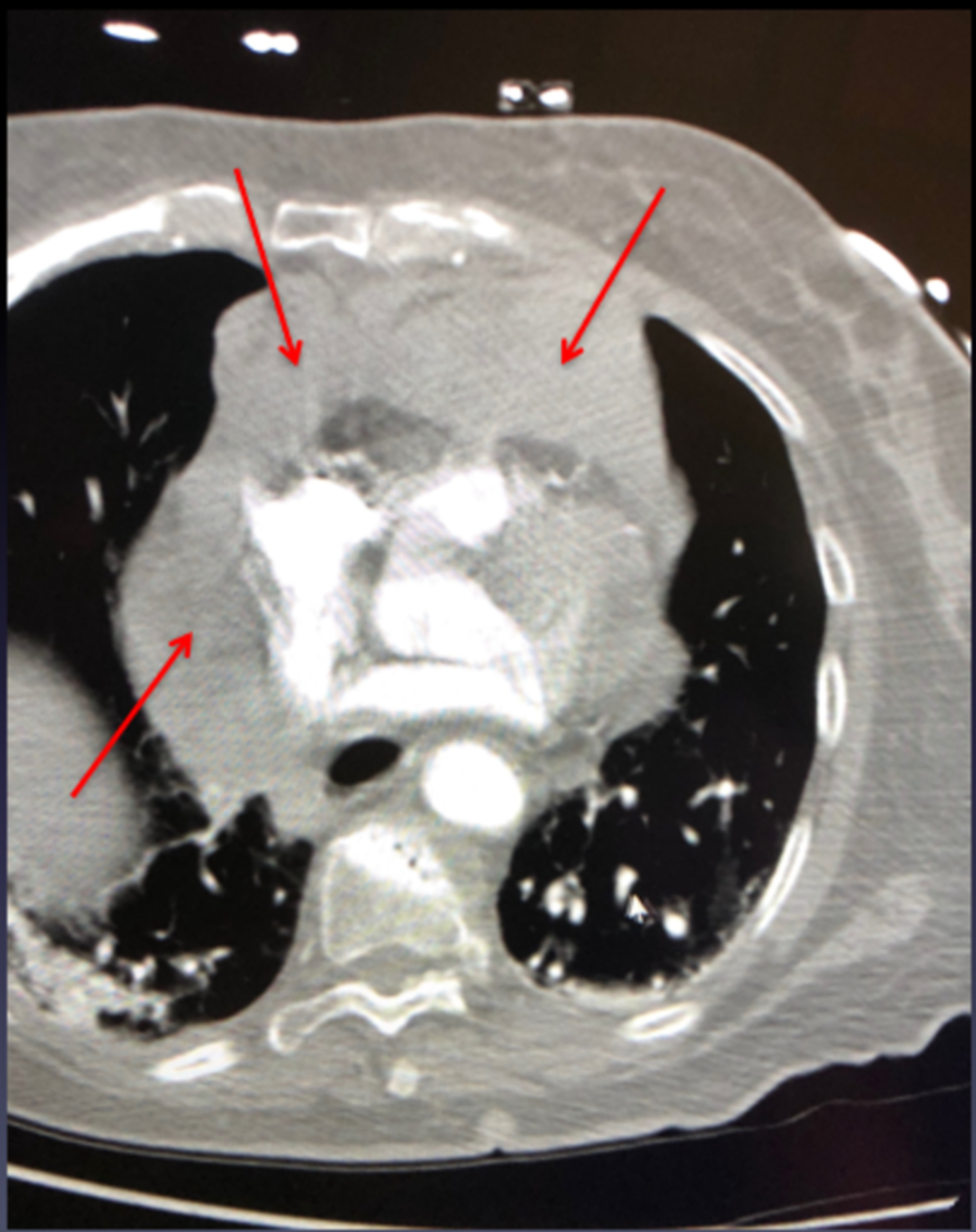
Lymph nodes enlargement with a maximum size of 13.63 mm (arrow) in the ...
Lymph-Node-Biopsy-Lymphoma
Sentinel-Lymph-Node-Biopsy-Melanoma
Lymph-Node-Biopsy-Scar
Neck-Lymph-Node-Biopsy-Procedure
Inguinal-Lymph-Node-Biopsy
Groin-Lymph-Node-Biopsy-Procedure
Benign-Lymph-Node
Lymph-Node-Removal
Fine-Needle-Biopsy-Lymph-Node
Lymph-Nodes-On-Mammogram
Cancer-Neck-Lymph-Nodes
Lymph-Node-Biopsy-Diagram
Lymph-Node-Removal-Surgery
Inflamed-Lymph-Nodes
Axillary-Sentinel-Lymph-Node
Axillary-Lymph-Nodes-Ultrasound