Please enter url.
Login
Logout
Please enter url.
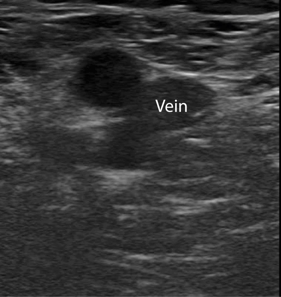
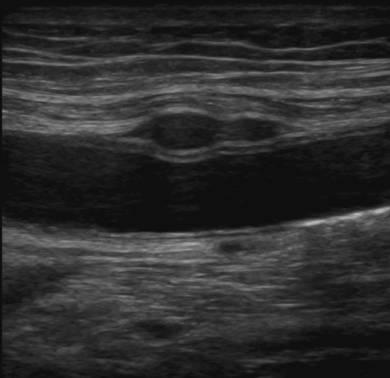
Venous Thrombosis Ultrasound
ar.inspiredpencil.com
source
Comments
Ultrasound image of a left axillary vasculature. | Download Scientific ...
Hints to ultrasound findings given by lab values? - Sononotes
Ultrasonographic image of a deep vein thrombosis ( | Deep vein ...
Magnetic resonance imaging of endometriosis: a common but often hidden ...
Venous duplex ultrasound image of thrombosed saphenofemoral junction ...
Pediatric and Adolescent Breast Masses: A Review of Pathophysiology ...
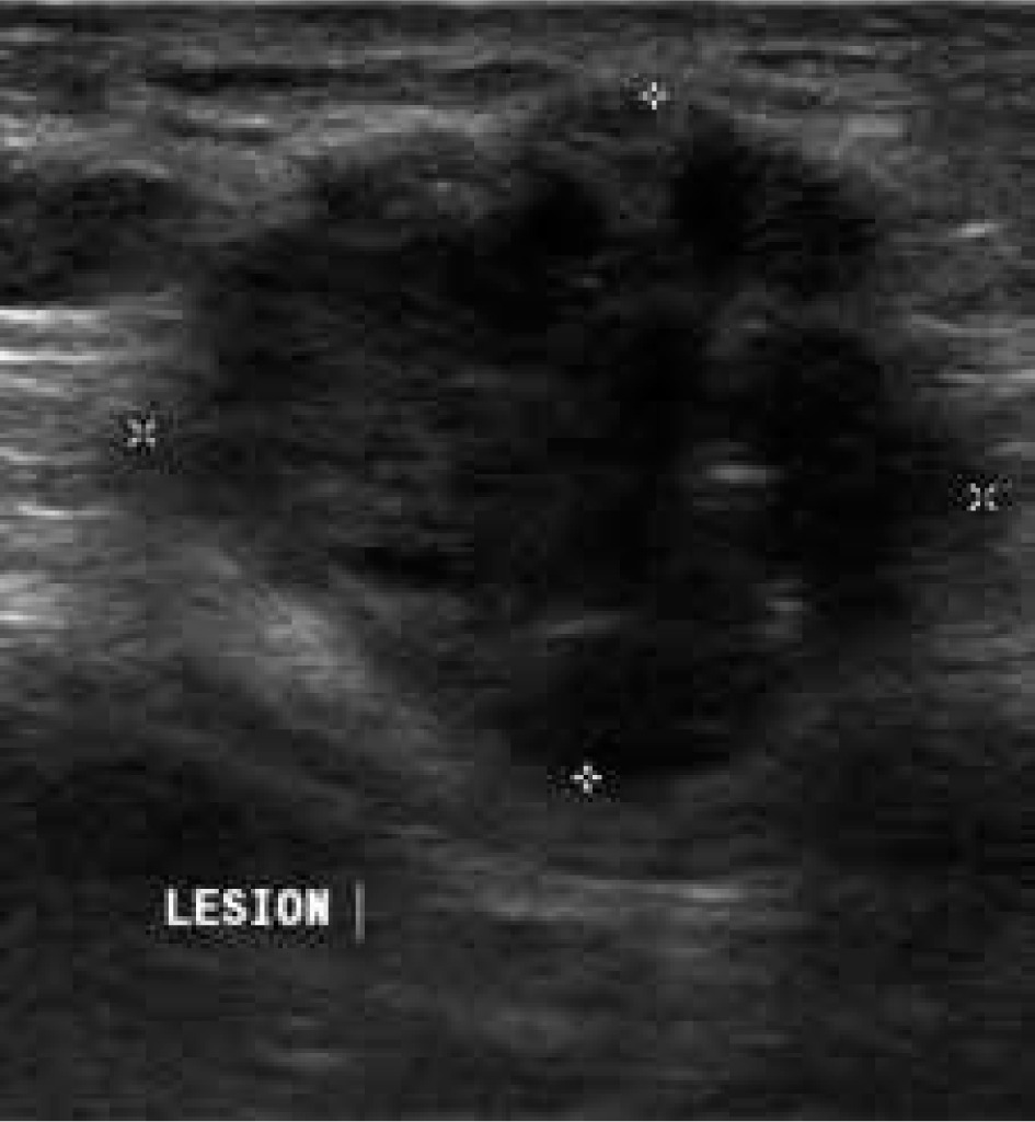
Breast and Axilla | 5.3 Pitfalls : Case 5.3.2 Benign lesions mimicking ...
Papillary carcinoma of thyroid. US and MRI features of U5 rare ...
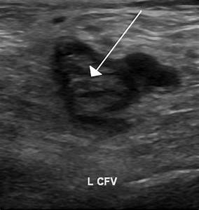
| Intraluminal mass. The persistent thrombus (arrow) originating from ...
(PDF) Life-Threatening, Bleeding Pseudoaneurysm of the External Iliac ...
Echogenic thrombus is visible in the right popliteal vein (vein on the ...
Ultrasound images showing the femoral vein (V) initially lying lateral ...
Pleural Effusion, Empyema, and Pneumothorax | Thoracic Key
Unilateral Leg Swelling Secondary to Venous Insufficiency | Thoracic Key
12 | Radiology Key
Epidermoid cyst - left testicular benign mass | Eurorad
Normal CT venography of the lower extremities. The venous system of the ...
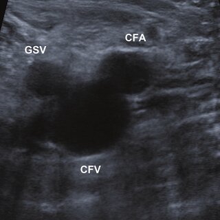
Transverse ultrasound image of the common femoral artery (CFA) and vein ...
Peripheral vessels | 10.1 Peripheral arteries : Case 10.1.3 Vasculitis ...
(PDF) A novel presentation of tubular adenoma of the breast as an ...
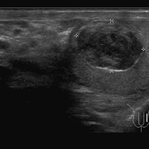
Gray-scale ultrasound image shows a well-circumscribed nodule ...
E2D ultrasound to identify the anatomy of the target vein. Image of the ...
Peripheral vessels | 10.1 Peripheral arteries : Case 10.1.3 Vasculitis ...
Hip and Thigh Ultrasound | Radiology Key
December 2013 | Ultrasound Cases
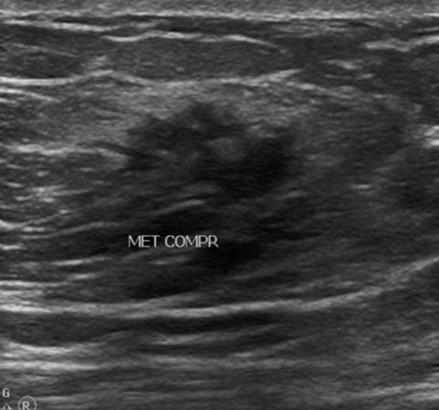
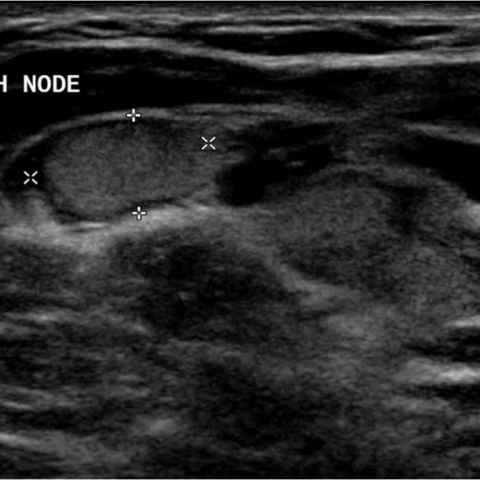
Low magnification shows metastatic papillary carcinoma of lymph node ...
(PDF) A Mammary Nodule Mimicking Breast Cancer
Typical image of mixed masses | Download Scientific Diagram
Imaging | Thoracic Key
Abdomen and retroperitoneum | 1.11 Abdominal wall : Case 1.11.5 ...
Several simple cysts present bilaterally, largest at 9 o'clock position ...
Peripheral vessels | 10.1 Peripheral arteries : Case 10.1.3 Vasculitis ...
Pyloric Stenosis – Undergraduate Diagnostic Imaging Fundamentals
Musculoskeletal Joints and Tendons | 6.1 Shoulder : Case 6.1.2 Biceps ...
Peripheral vessels | 10.1 Peripheral arteries : Case 10.1.3 Vasculitis ...
Positive-DVT-Ultrasound
Popliteal-Vein-Ultrasound
Compression-Ultrasound-DVT
DVT-Ultrasound-Leg
Ultrasound-for-DVT
Doppler-Ultrasound-DVT
Duplex-Ultrasound-DVT
DVT-Ultrasound-Anatomy
Venous-Duplex-Ultrasound-DVT
Lower-Extremity-DVT-Ultrasound
Acute-DVT-Ultrasound
Chronic-DVT-Ultrasound
Leg-Veins-Ultrasound
Basilic-Vein-Ultrasound
Soleal-Vein-DVT-Ultrasound
Superficial-Femoral-Vein-DVT