Please enter url.
Login
Logout
Please enter url.
[PDF] Bilateral perisylvian infarct: a rare cause and a rare occurrence ...
semanticscholar.org
source
Comments
Desensitization to trimethoprim-sulfamethoxazole in a toxoplasmic ...
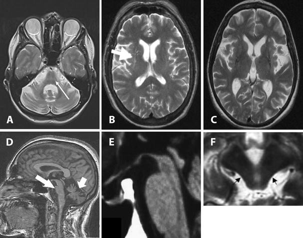
MRI at diagnosis and follow-up. (A, B) Axial T2-weighted (T2WI) and ...
Initial MR images in two subjects. Top row shows initial T1 SE post ...
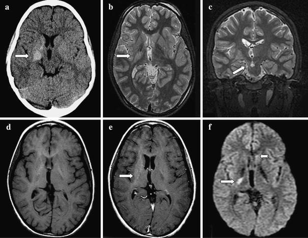
Patient 1. Axial non-contrast-enhanced CT brain scan revealing a left ...
COVID-19-related intracranial imaging findings: a large single-centre ...
Visual and neurologic sequelae of methanol poisoning in Saudi Arabia ...
Magnetic resonance imaging results. Notes: Hyperintensities noted in ...
Magnetic resonance imaging for the diagnosis of Parkinson’s disease ...
Case 3-2010 — A 5-Month-Old Boy with Developmental Delay and ...
Acute encephalitis with refractory, repetitive partial seizures - Brain ...
Cerebral MRI abnormalities associated with vigabatrin therapy - Pearl ...
Pneumocephalus. This 58-year-old woman underwent bilateral VIM DBS for ...
| The distributions of symptoms and juxtacortical small lesions ...
Inherent diagnostic and treatment challenges in germinoma of the basal ...
Patient with CSF analysis positive for meningitis A: Axial FLAIR ...
Vitamin B12 Deficiency With Bilateral Globus Pallidus Abnormalities ...
MRI Shows Pediatric Stroke Case Associated with COVID-19 | Diagnostic ...
Heterogeneity in age-related white matter changes | SpringerLink
Brain MRI findings in children with Leigh-like syndrome. a Brain MRI ...
Radiographic resolution of an acute silent cerebral ischemic event ...
(PDF) Autoimmune Encephalitis: Pathophysiology and Imaging Review of an ...
Anticoagulation Therapy and Imaging in Neonates With a Unilateral ...
The protean manifestations of central nervous system IgG4-related ...
Cerebral MRI abnormalities associated with vigabatrin therapy - Pearl ...
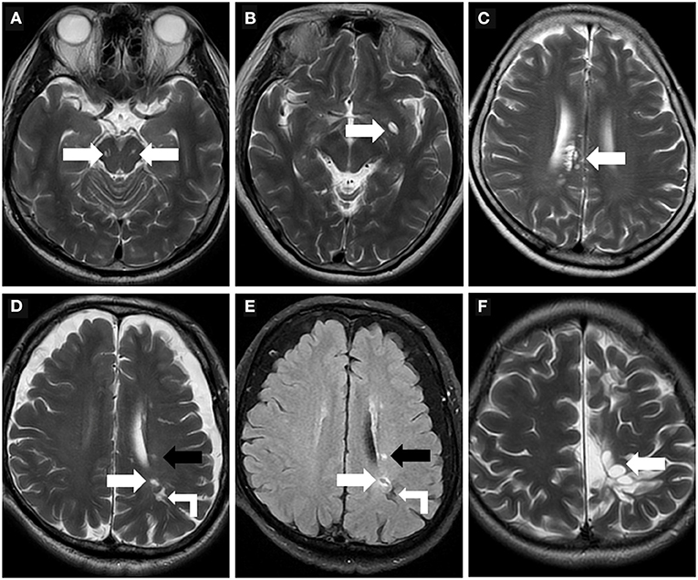
Frontiers | Perivascular Spaces, Glymphatic System and MR
Diagnostic Criteria of Acute Necrotizing Encephalopathy Proposed by ...
Dat Scan For Parkinson's Disease Procedure - ParkinsonsInfoClub.com
High-resolution, T2-weighted, FSE MRI (voxels of 0.47 mm ϫ 0.47 mm ϫ 2 ...
CAA. A, Axial brain CT scan shows a subtle left rolandic... | Download ...
Forty-year-old male with diffuse post-hypoxic demyelination (one month ...
Magnetic resonance imaging (MRI) at presentation. a T 2 w sequence ...
A common pattern of brain MRI imaging in mitochondrial diseases with ...
Venous complications following petrosal vein sectioning in surgery of ...
Cerebrovascular disease in pregnancy and puerperium: perspectives from ...
Patient 4. Apert syndrome. A , Axial spin-echo (3000/120) MR image ...