Please enter url.
Login
Logout
Please enter url.
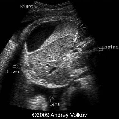
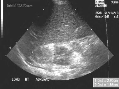
Adrenal Gland On Ultrasound
mavink.com
source
Comments
a, b. Standard ultrasound – hypoechoic focal lesion in the middle part ...
The Adrenal Glands | Obgyn Key
Neonatal Neurosonography | Radiology Key
Right Flank Pain
Consolidated lung in a mechanically ventilated patient with ...
(PDF) Novel variants in COL4A4 and COL4A5 are rare causes of FSGS in ...
2D ultrasonogram in sagittal plane of right kidney in a 9-year-old ...
Renal Failure | Radiology Key
Sonography image showing a 7.9-cm mass with free fluid [←] around it ...
📃 Subdiaphragmatic abscess and jejunal atresia
Aspect évolutif d'un abcès amibien uniloculé des segments V et VI ...
Station 4R (right paratracheal) lymph node, described as oval shape ...
Ultrasonogram of the abomasum in a camel. Image was taken in the right ...
Regular (a) and irregular (b) endometrial-myometrial border. In (b ...
Marked abdominal distension and presentation of Cullen's sign (edema ...
Adrenal Hemorrhage Imaging and Diagnosis: Practice Essentials ...
Subcarinal lymphadenopathy (station 7) Visualized via the esophagus ...
The levels of total bilirubin (T-Bil), direct bilirubin (D-Bil ...
(A) A transverse ultrasonography of the liver showed coarse ...
Homogeneous (a) and heterogeneous (b) internal endometrial echogenicity ...
Neonatal Head images Flashcards | Quizlet
Longitudinal ultrasound of the right upper quadrant showing crescentic ...
Axial transcranial color Doppler sonography shows M1 segment of the MCA ...
Nontranslobar lung consolidation. Longitudinal scan at the PLAPS point ...
(PDF) Bilateral cavernous sinus syndrome in dogs: 6 cases (1999-2004)
Transverse US through the liver shows the classic “starry night ...
6 (a) Hemangioma in the dorsal chest wall. Soft swelling on the left ...
Small Animal Abdominal Ultrasonography, Part 1: A Tour of the Abdomen ...
Transverse scan of the right hepatic lobe shows a coarse reticular ...
Abdominal Ultrasound Registry 2012 Flash Cards Flashcards - Cram.com
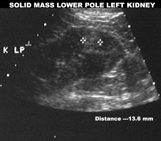
Renal Cell Carcinoma
(A,B) Minor patchy echogenicity (white arrows) on the ultrasound scans ...
Endometriosis: Radiologic-Pathologic Correlation | RadioGraphics
1. Ultrasound with moderate hidronefrosis. | Download Scientific Diagram
In the right lobe, ultrasound showed 2 nodular lesions, with similar ...
Mass-On-Adrenal-Gland
Adrenal-Gland-Adenoma
Adrenal-CT-Scan
Enlarged-Adrenal-Gland
Growth-On-Adrenal-Gland
Adrenal-Gland-Pain
Thyroid-and-Adrenal-Gland
Cancer-of-Adrenal-Gland
Symptoms-of-Adrenal-Tumor
Adrenal-Gland-Pheochromocytoma
Nodule-On-Adrenal-Gland
Adrenal-Gland-Cyst
Signs-of-Adrenal-Tumor
Left-Adrenal-Mass
Kidney-and-Adrenal-Gland
Adrenal-Gland-Disease


![Sonography image showing a 7.9-cm mass with free fluid [←] around it ...](https://www.researchgate.net/publication/278160096/figure/fig1/AS:272550001377313@1441992392982/Sonography-image-showing-a-79-cm-mass-with-free-fluid-around-it_Q320.jpg)