Please enter url.
Login
Logout
Please enter url.
Basal Ganglia Anatomy Ct
mavink.com
source
Comments
| Bilateral basal ganglia necrosis with T2w hyperintense alterations ...
Comparison of MRI and CT for Detection of Acute Intracerebral ...
Axial T2 ( ) and fluid-attenuated inversion recovery ( ) images show ...
Vigabatrin‐associated reversible MRI signal changes in patients with ...
Postpartum angiopathy. A 23-year-old primigravida presented with ...
(A) Axial T1-weighted magnetic resonance (MR) image showing ...
(PDF) Reto diagnóstico y terapéutico en la hemorragia intracerebral por ...
Figure 1 from Posterior Reversible Encephalopathy Syndrome With ...
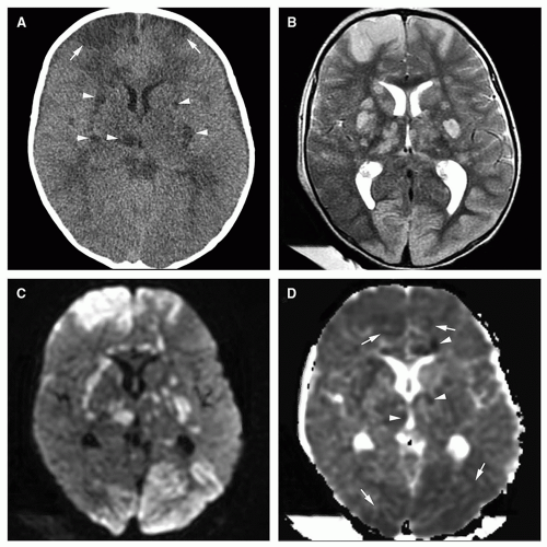
Axial diffusion-weighted imaging magnetic resonance imaging (MRI) of ...
Diagrammatic illustration of the right medial temporal anatomy and the ...
Infections of the Developing and Mature Nervous System | Radiology Key
Magnetic resonance imaging for the diagnosis of Parkinson’s disease ...
Axially oriented T 2-weighted MRI images showing extensive damage to ...
MRI of neonate affected by herpes virus type 1 (A-D) presenting with ...
Patient 2. MRI FLAIR (a) showing hyperdensity in the right thalamus ...
A (MRI in T2 FLAIR sequence) shows multiple ischemic lesions in ...
(A) T1, (B) T2, (C) diffusion weighted imaging, (D) susceptibility ...
(A) CT Scan and (B) T2 Flair MRI: Cerebral imaging showing large ...
A Case of Early-Onset Rapidly Progressive Dementia | Cerebrovascular ...
Brain MRI showing right frontoparietal lobe cortical swelling with ...
Frequency of MELAS main mutation in a phenotype-targeted young ischemic ...
| Computer tomography showing hypodense areas bilaterally in the ...
Gram staining of the cerebrospinal fluid sample showing short ...
Diagnostics | Free Full-Text | Cerebral Infarction and Evan’s Ratio on ...
| Detection of axonal injury with conventional magnetic resonance ...
The sixth month aged male boy with diagnosed thrombosis at torcular ...
CJD showing sharp wave complexes [9]. | Download Scientific Diagram
MRI findings at the age of 1 year (A), 13 years and 10 months (B, C ...
Brain MRIs. (A) An initial brain MRI in a coronal T2-weighted sequence ...
Post-mortem MRI versus conventional autopsy in fetuses and children: a ...
Post-ictal EEG showed focal paroxysmal epileptiform sharp and slow ...
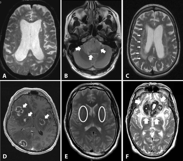
Representative MR images showing tumor necrosis, rim enhancement ...
MRIs of CNTNAP1 patients and controls | Download Scientific Diagram
Community-acquired bacterial meningitis - The Lancet
Laura BENJAMIN | Principal Clinical Research Fellow | MRCP PhD ...
Basal-Ganglia-in-CT
Basal-Ganglia-Axial-MRI
Calcification-of-the-Basal-Ganglia
Basal-Ganglia-CT-Scan
Basal-Ganglia-Coronal-Section
Basal-Nuclei
Basal-Ganglia-Brain-MRI
Basal-Ganglia-Infarct
Basal-Ganglia-Stroke-CT
Basal-Ganglia-Infarction
Basal-Ganglia-Damage
Brain-Anatomy-Basal-Ganglia
Striatum-Basal-Ganglia
Left-Basal-Ganglia-Infarct
Basal-Ganglia-Brain-Cross-Section
Basal-Ganglia-Brain-Functions


![CJD showing sharp wave complexes [9]. | Download Scientific Diagram](https://www.researchgate.net/publication/363182144/figure/fig4/AS:11431281082463034@1662038503012/Pattern-of-cortical-changes-in-CJD-9_Q640.jpg)


