Please enter url.
Login
Logout
Please enter url.
Quantitative EEG for the detection of brain ischemia | Critical Care ...
ccforum.biomedcentral.com
source
Comments
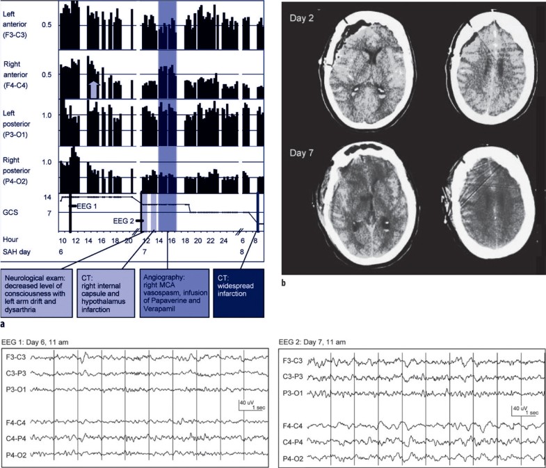
Alpha/delta ratio (ADR) calculated every 15 min and Glasgow Coma Score ...
Non-convulsive Status Epilepticus and Non-convulsive Seizures in ...
(A) From left to right; DWI-ADC shows diffusion restriction in the ...
Right mesial temporal sclerosis (MTS) in Patient n. 9 | Download ...
(PDF) Multimodal imaging in nonlesional medically intractable focal ...
Amplitude-Integrated EEG and Its Potential Role in Augmenting ...
Hemodynamic response patterns in a patient with childhood epilepsy with ...
EEG and imaging findings predicting poor neurologic outcome after ...
Epilepsy Surgery in Infants and Children | Neupsy Key
Insular Epilepsy: Semiology and Noninvasive Investigations : Journal of ...
(PDF) Technical Aspects of SEEG and Its Interpretation in the ...
Factors Associated With Risk of Recurrent Transient Global Amnesia ...
(PDF) Visualizing prolonged hyperperfusion in post-stroke epilepsy ...
New-onset tic disorder following circumscribed brain injury - Journal ...
(a) Generalized 3-Hz spike-wave discharges during absence seizures are ...
( a ) Axial FLAIR and ( b ) sagittal and ( c ) coronal T1-weighted MR ...
The Treatment of Intracranial Hypertension. Algorithm of Treatment and ...
Which EEG patterns in coma are nonconvulsive status epilepticus ...
(PDF) Magnetic Resonance Spectroscopy for Evaluating Portal-Systemic ...
Relationships between the epileptic focus and hand area in central ...
Example of focal hypoperfusion seen on sASL in a 33-year-old right ...
Figure 1 from Early Modified Functional Hemispherectomy in a Young ...
Intracranial Pressure Fluctuations Mimicking Seizures in a P ...
Caso 2. Esclerosis mesial temporal derecha (A). Lobectomía temporal ...
The bipolar montages (double banana) of electroencephalography (EEG ...
Development over time of the aEEG background between days 1 and 3 after ...
Example of amplitude-integrated electroencephalogram (aEEG)/EEG ...
EEG recordings showed that the interictal EEG background was normal ...
John Libbey Eurotext - Epileptic Disorders - Concept of epilepsy ...
Estimated direction of propagation of epileptiform activity along ...
Audiological and radiological findings A : Pure tone audiometry (PTA ...
Surgical treatment of two patients with infantile spasms in early ...
(A) Ictal EEG onset and interictal sharp wave in patient 8. The center ...
(A, B) Case 1. (A) Axial T2 MRI showing changes consistent with remote ...
EEG picture (above) and brain (below) of patient 2. Note the epileptic ...
Stroke-EEG
Hemorrhagic-Brain-Stroke
Hemorrhagic-Stroke-CT
Hemorrhagic-Stroke-CT-Scan
Hemorrhagic-Stroke-Pathophysiology
Ischemic-vs-Hemorrhagic-Stroke
What-Is-a-Hemorrhagic-Stroke
Hemorrhagic-Stroke-Types
2-Types-of-Hemorrhagic-Stroke
Seizure-On-EEG
EEG-Symptoms
Hemorrhagic-Stroke-Anatomy
Abnormal-EEG
EEG-Waves-Stroke
EEG-After-Stroke
Stroke-EMG