Please enter url.
Login
Logout
Please enter url.
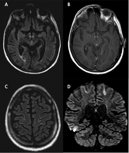
Cortical Linear Lesions in Wernicke’s Encephalopathy: Can Diffusion ...
neuro.psychiatryonline.org
source
Comments
Cortical Linear Lesions in Wernicke’s Encephalopathy: Can Diffusion ...
EPOS™
Chapter 21 – Cervical Artery Dissection and Cerebral Vasculitis ...
EPOS™Primary CNS Lymphoma is identified as a CT hyperdense enhancing ...
Treatment of severe acute necrotizing encephalopathy of childhood with ...
Pyogenic abscess. Axial FLAIR (A) demonstrates a round hypointense mass ...
Repeat brain MRI after 44 months: axial FLAIR images (reconstructed ...
Cortical laminar necrosis related to migrainous cerebral infarction
Teaching NeuroImages: Transient mutism associated with splenium lesion ...
Cavitation After Acute Symptomatic Lacunar Stroke Depends on Time ...
MRI findings of case 1. Diffusion-weighted image (a) and T2- weighted ...
Comparative effects of Micruroides euryxanthus, Micrurus fulvius ...
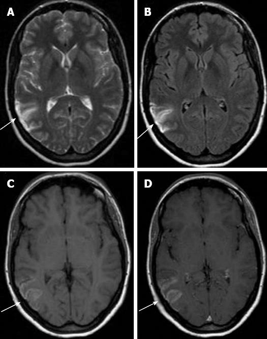
Focal Status Epilepticus-Related Unilateral Brain Edema: Magnetic ...
Delayed neurological deficits after endovascular placement of a ...
Patient With Voltage-Gated Potassium-Channel (VGKC) Limbic Encephalitis ...
Sporadic Creutzfeldt-Jakob disease: A probable diagnosis based on ...
Iatrogenic Coagulopathy and the Development of Posterior Reversible ...
Axial MRI Brain of Case 2 demonstrating T2-FLAIR hyperintensities ...
Figure 1 from Mild encephalitis/encephalopathy with a reversible ...
Patient #14. MRI axial diffusion (A), ADC map (B), axial FLAIR (C), and ...
AStepAway - New Search Experience | Carbon monoxide poisoning, Mri ...
Fatal metabolic stroke in a child with propionic acidemia 11 years post ...
Brain Sciences | Free Full-Text | New Insight in Hyperinsulinism ...
Diffusion‐weighted MRI abnormalities in an outbreak of Streptococcus ...
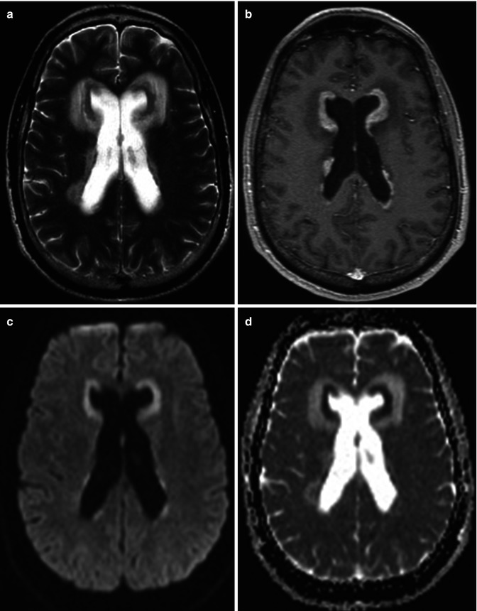
Frontiers | Progressive Multifocal Leukoencephalopathy Diagnosed by ...
Axial FLAIR -hyperintense white matter bilateral temporo-occipital (A ...
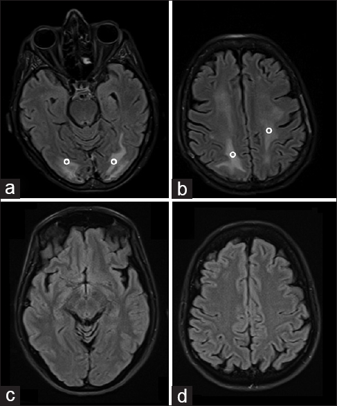
Claustrum hyperintensities: A potential clue to autoimmune epilepsy ...
Metastasis and Other Tumors of the CNS | Radiology Key
Imaging and CSF analyses effectively distinguish CJD from its mimics ...
Rhinocerebral Mucormycosis with Top of Basilar Artery Syndrome ...
The MRI was performed on the 15th postoperative day, showing the state ...
Three-year-old male (patient 4) with hyperacute thrombosis (hours to ...
Posttransplant lymphoproliferative disease (PTLD): a patient with a ...
Late Onset of Psychotic Symptoms in a Patient With Cavum Septum ...
HIV encephalitis. Diffuse range of FLAIR hypersignal and T2 of ...