Please enter url.
Login
Logout
Please enter url.
Sacroiliac Joint Dysfunction Mri
ar.inspiredpencil.com
source
Comments
MR Imaging–based Assessment of the Female Pelvic Floor | RadioGraphics
An 8-month-old with inverted left anterior labrum. PD FS axial image ...
Sacral insufficiency fracture in a non-athlete young female
CT-/X-Ray-Guided Technique in Sacral Fusion | Neupsy Key
EPOS™
Využitie MRI vyšetrenia pri diagnostike axiálnej spondylartritídy ...
Frontiers | Axial Spondyloarthritis: Mimics and Pitfalls of Imaging ...
Coronal MR of the sacrum. A Bone lesion with T1WI hypointensity at left ...
Frontiers | The Role of Imaging in Diagnosing Axial Spondyloarthritis
MRI findings of active inflammation and chronic structural damage in ...
Comparison of fat-saturated T2-weighted and contrast-enhanced fat ...
MRI of superficial inguinal lymph nodes and site of inoculation ...
On MR images taken at 7 weeks postpartum, the bone marrow edema in the ...
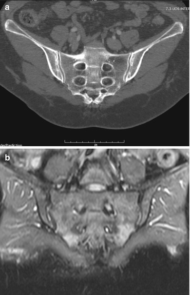
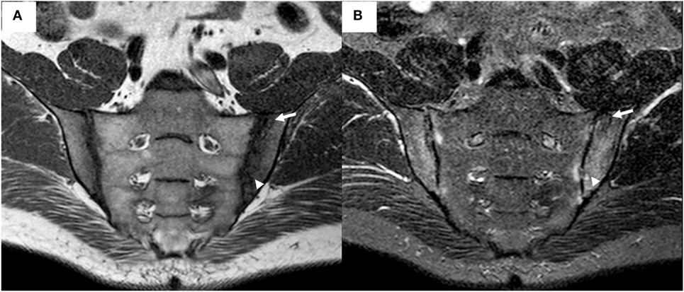
35-year-old female with dedicated Sacroiliac joint MRI and CT. (A) MRI ...
(PDF) Percutaneous transgluteal computed tomography-guided aspiration ...
-A, B -Coronal T1 sacroiliac joint magnetic resonance imaging (MRI ...
Knee arthritis in psoriatic arthritis. Sagittal ( a ) and axial ( b ...
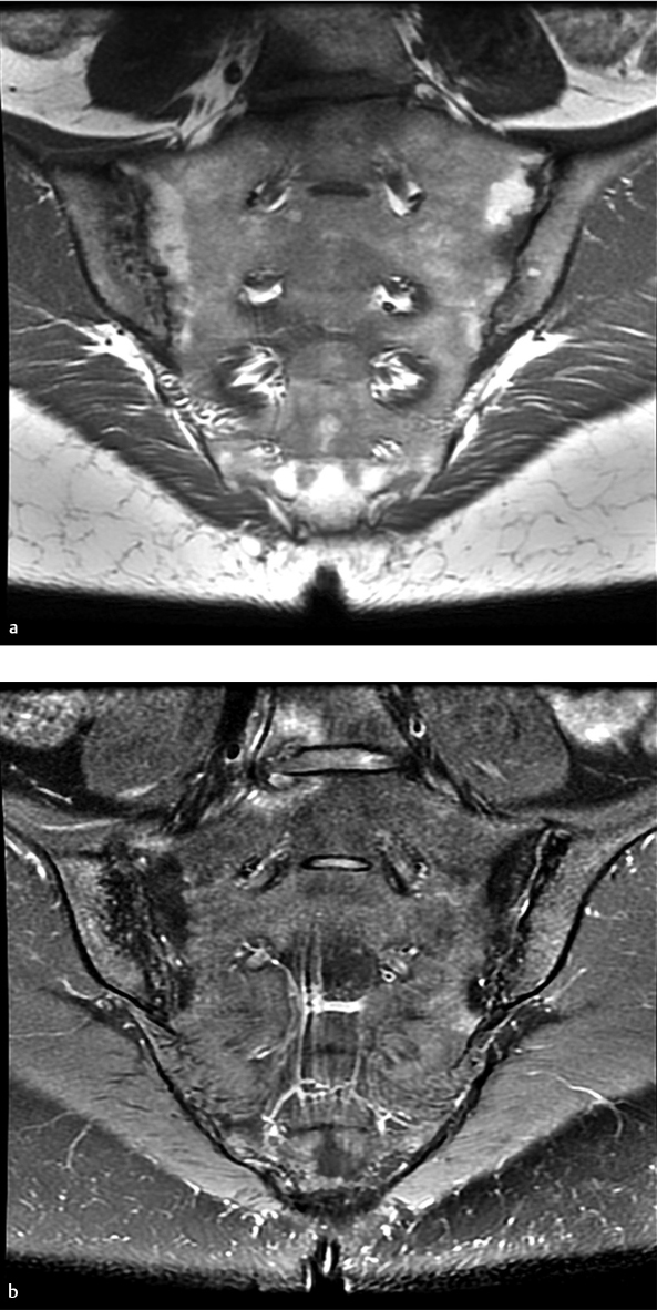
Oedema of sacroiliac joint at different sections of magnetic rezonans ...
Morphology of the most relevant lesion types on sacroiliac joint MRI in ...
(A) Lumbar-spine magnetic resonance imaging (MRI) (coronal view) shows ...
JCM | Free Full-Text | Diagnostics of Sacroiliac Joint Differentials to ...
a T2-weighted axial MRI demonstrating lateralized (left) and medialized ...
JCM | Free Full-Text | Diagnostics of Sacroiliac Joint Differentials to ...
(PDF) Symptomatic Lumbosacral Transitional Vertebrae (Bertolotti ...
Endoscopic Repair of Full-Thickness Gluteus Medius and Minimus Tears ...
MRI of the lumbar spine demonstrates a large disk herniation on level ...
spa-imaging.org
Part 5: Arthropathies | Radiology Key
Osteitis Condensans Ileii. T1 and STIR MRI sequences showing iliac ...
A 41 year-old woman with chronic sacroiliitis represented by multiple ...
(A) T1-weighted magnetic resonance imaging (MRI), showing no definite ...
On enhanced T1W lumbar axial MRI, paraspinal (black arrows) and psoas ...
Radiologia Brasileira - Espondiloartropatias: critérios de ressonância ...
JCM | Free Full-Text | Diagnostics of Sacroiliac Joint Differentials to ...
A 40-year-old female patient with a diagnosis of nr-axSpA well ...
Ankylosing-Spondylitis-Spine-MRI
Right-SI-Joint
Normal-SI-Joint-MRI
Sacrum-MRI
Sacroiliac-Joint-Inflammation
SI-Joint-Fusion-Surgery
Sacroiliac-Joint-Sclerosis
SI-Joint-Anatomy
Sacroiliitis-MRI
SI-Joint-Syndrome
SI-Joint-CT-Scan
Diagram-of-SI-Joint
SI-Joint-Arthritis
Sacroiliac-Si-Joint-Pain
SI-Joint-Instability
Sternoclavicular-Joint-MRI