Please enter url.
Login
Logout
Please enter url.
An Epidemiological Study on Paediatric Brain MRIs with a Focus on ...
emjreviews.com
source
Comments
An Epidemiological Study on Paediatric Brain MRIs with a Focus on ...
Brush Sign Is Associated With Increased Severity in Cerebral Venous ...
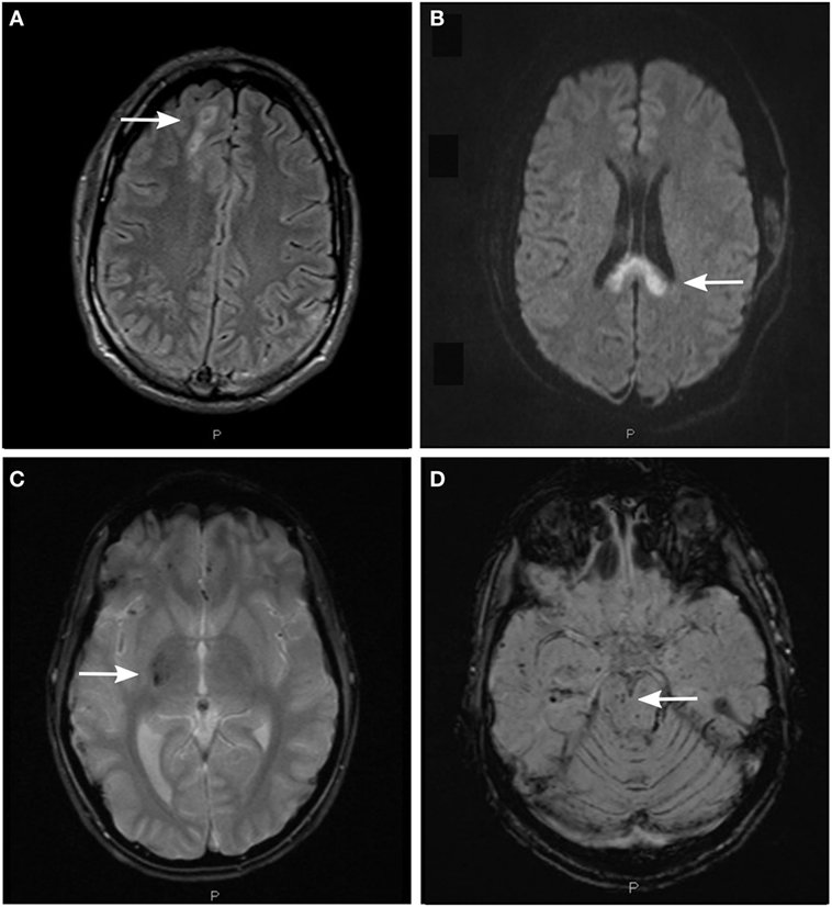
(A,B) A hyperintense lesion within the right caudate nuclei is observed ...
Periventricular leukomalacia on conventional and wave-CAIPI MPRAGE. A ...
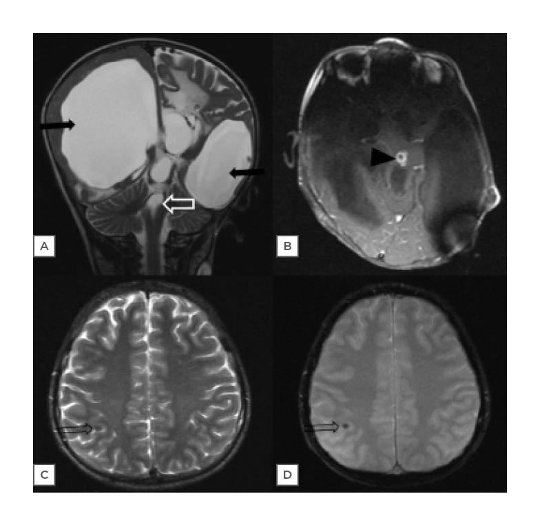
A child with imaging features of tuberous sclerosis and SWS. a Axial ...
Brain MRI roulette | Practical Neurology
Ictal urge to defecate associated with a right-sided mesial temporal ...
Frontiers | Mitochondrial Neurogastrointestinal Encephalomyopathy ...
Cureus | Flaccid Brachial Monoplegia As Initial Presentation in a ...
Marchiafava–Bignami disease-like lesions due to central nervous system ...
Magnetic resonance imaging of brain showing high T2 signal in bilateral ...
Isolated neonatal MRI punctate white matter lesions in very preterm ...
Cranial MRIs. A, Enhanced MRI scan performed on April 01, 2019 with ...
Perinatal Stroke | Obgyn Key
| (A) Axial T1-weighted image showing bilateral subdural hemorrhages ...
Intracranial hemorrhagic manifestations in adenosine deaminase 2 ...
Mild malformation of cortical development with oligodendroglial ...
Raised Plasma Neurofilament Light Protein Levels Are Associated with ...
MRI of the brain and spine of a 34-year-old female patient with ...
Frontiers | Current Opportunities for Clinical Monitoring of Axonal ...
A 3-month-old boy with abusive head trauma. a An axial... | Download ...
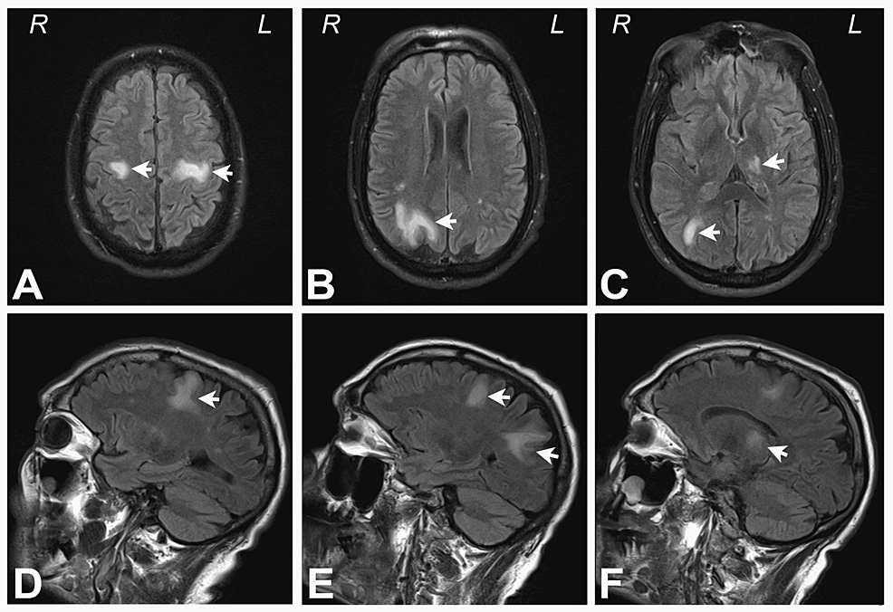
Multiple patchy hyperintensity on T2 Flair (a and b: arrows) were found ...
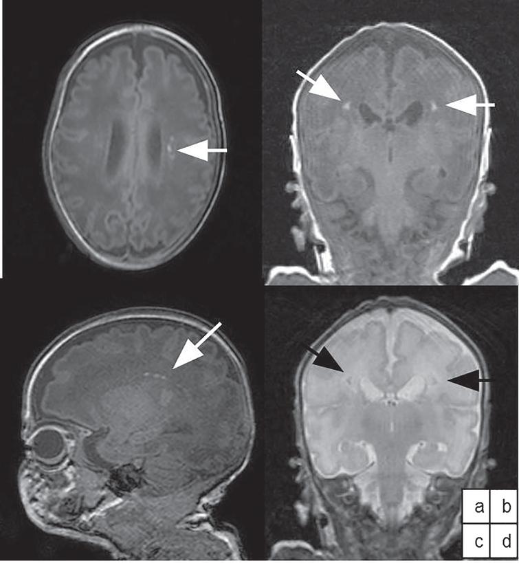
(ID 109): Dysmorphic left lateral ventricles and periventricular ...
Measuring Permeability in Acute Ischemic Stroke - Neuroimaging Clinics
Diffuse cauda equina enhancement in a middle aged male with Susac ...
Bone Cancer Skull Xray - CancerWalls
Brain magnetic resonance imaging (MRI) of a 25-day-old neonate with ...
Histopathology and immunohistochemistry of the right posterior parietal ...
Lissencephaly Radiology
Figure 1 from Hypertensive slit ventricle syndrome: pseudotumor cerebri ...
Figure 1 from Response to “Do not miss Bickerstaff encephalitis as a ...
Parenchymal brain laceration with a fluid level. Case 18. A, Axial NCCT ...
enhanced Mri of a 6 mm slice at the start of the treatment with MPa ...
Location of hypertrophic pachymeningitis | Download Scientific Diagram
Brain MRI of subject P1. Representative images of cerebral MRI ...