Please enter url.
Login
Logout
Please enter url.
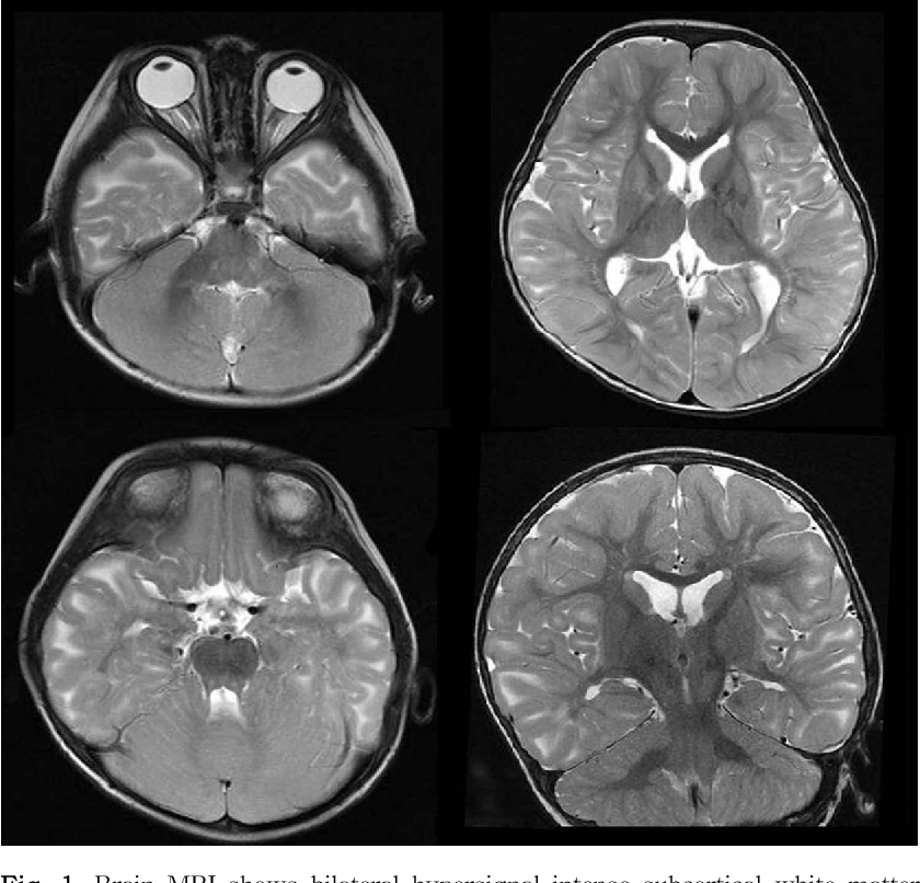
Cerebral MRI axial T2 weighted sequences (A–C) showing hyper T2 signal ...
researchgate.net
source
Comments
Xq26.1‐26.2 gain identified on array comparative genomic hybridization ...
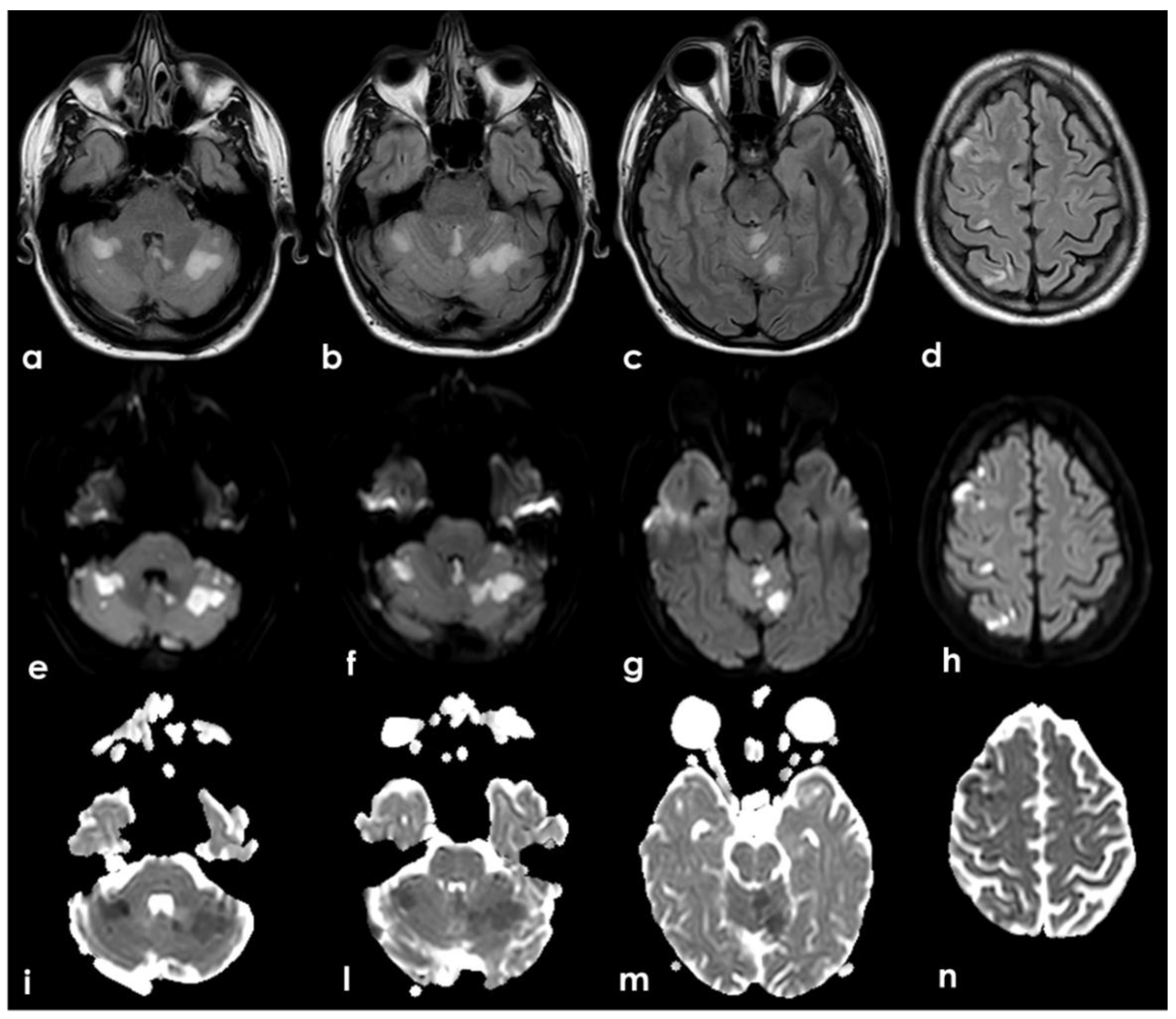
Head MRI results of the patient. ①–⑥ was on admission, and ⑦–⑨ was 1 ...
Brain MRI findings in a patient with non-neoplastic radiation induced ...
Intracranial involvement in acute invasive fungal rhinosinusitis ...
Brain MRI taken 16 days after spinal surgery shows small sites of acute ...
Case 5. A: Axial T2-weighted MR image showing a right frontal DLGG in a ...
Acute Pediatric Encephalitis Neuroimaging: Single-Institution Series as ...
Figure 1 from Intracerebral hemorrhage due to developmental venous ...
JCM | Free Full-Text | Neurovascular Manifestations of Iron-Deficient ...
mrcs-7-9-001-g001
Elderly woman with psychosis and unsteadiness | Practical Neurology
pediatric neurology: Predominant area of brain lesions in neonates with ...
"Two lumen" sign on brain MRI, 1 is in crescent shape: (A) T1 flair ...
Intracranial Meningeal Involvement in Churg-Strauss Syndrome | American ...
Neuroimaging data 12 days after the stroke. (a) Axial DWI showing ...
Panthotenate kinase-associated neurodegeneration and phospholipase A2 ...
Case of Aicardi-Goutieres Syndrome - Page 2
The transfrontal isthmus approach for insular glioma surgery in ...
Severe cerebral involvement due to idiopathic systemic capillary leak ...
Imaging of Brain Tumors | Radiology Key
Neuroradiologic Phenotyping of Galactosemia: From the Neonatal Form to ...
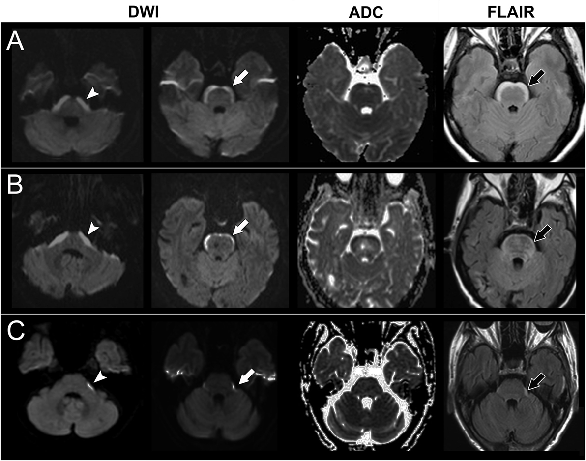
Frontiers | Novel Anterior Brainstem Magnetic Resonance Imaging ...
Teaching NeuroImages: Reversible paradoxical lithium neurotoxicity ...
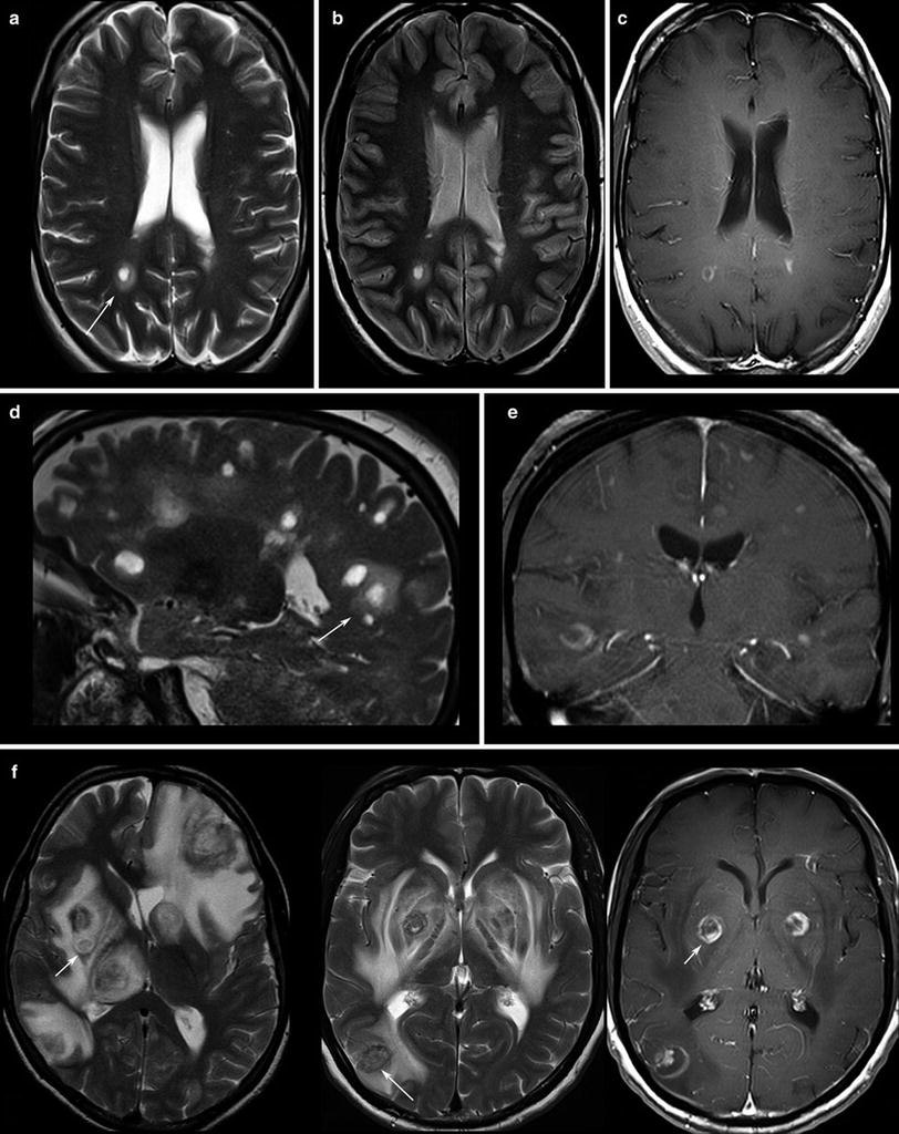
MRI FLAIR sequence axial cuts (a, b) and ADC map (c) showing bilateral ...
Chasing the wrong dragon: A new presentation of heroin-induced toxic ...
(PDF) Diagnostic Approach to Macrocephaly in Children
'Central' pachygyria with p.R264C TUBA1A mutations. This figure shows ...
Imaging Patterns Characterizing Mitochondrial Leukodystrophies ...
a. Axial head CT discloses obstructive hydrocephalus with hemorrhage ...
a Initial axial FLAIR images demonstrate marked high-signal intensity ...
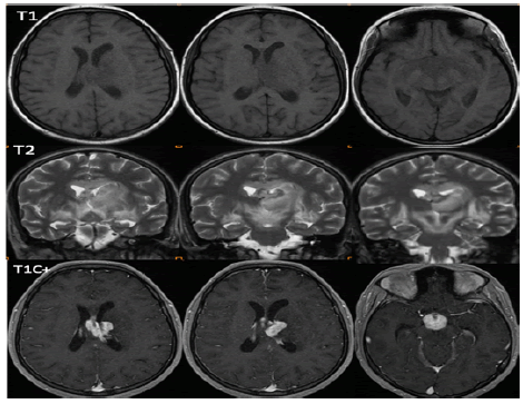
MRI studies, including a T1Gd and b FLAIR sequences, of patient #1 ...
Figure 1 from A case of megalencephalic leukoencephalopathy with ...
Risk factors for unchanged ventricles during pediatric shunt ...
Relapsing-Remitting Severe Bickerstaff's Brainstem Encephalitis - Case ...
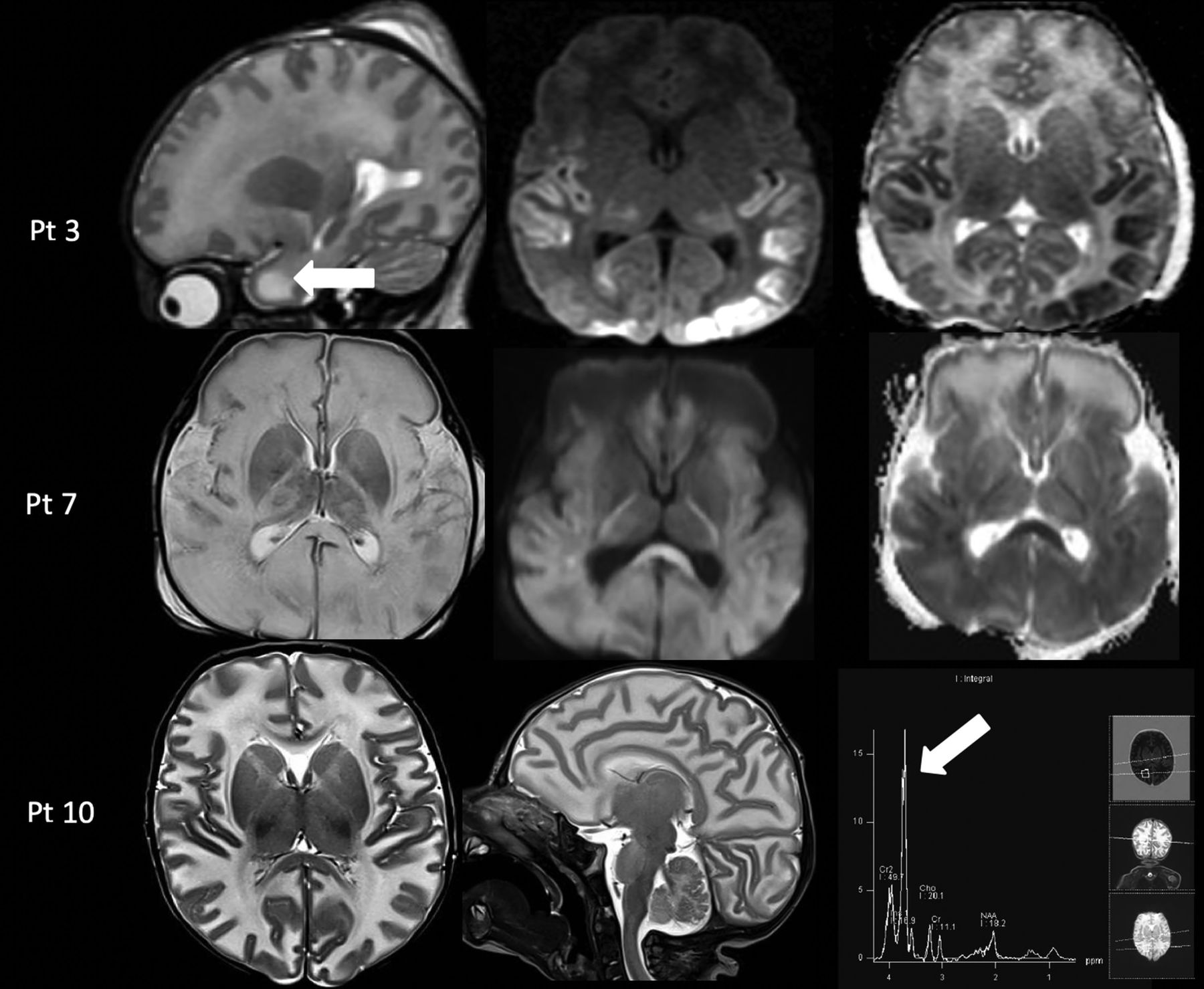
Brain MRI findings of 16 patients with cortical blindness. All patient ...