Please enter url.
Login
Logout
Please enter url.
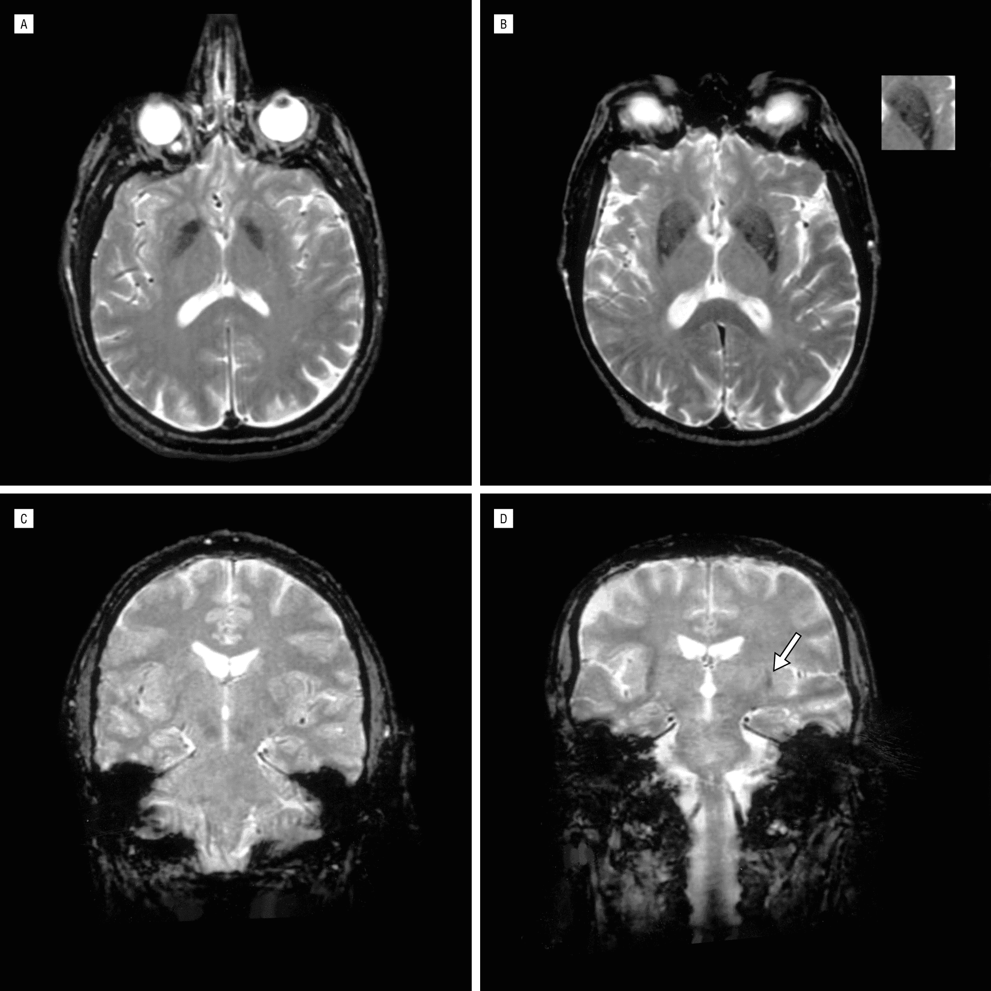
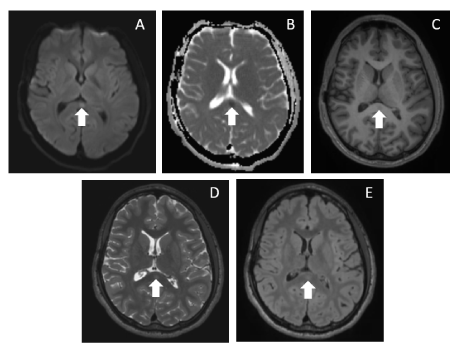
Follow up cranial MRI. The follow up cranial MRI showed no lesion on ...
researchgate.net
source
Comments
Information of 29 adult-onset MERS cases | Download Table
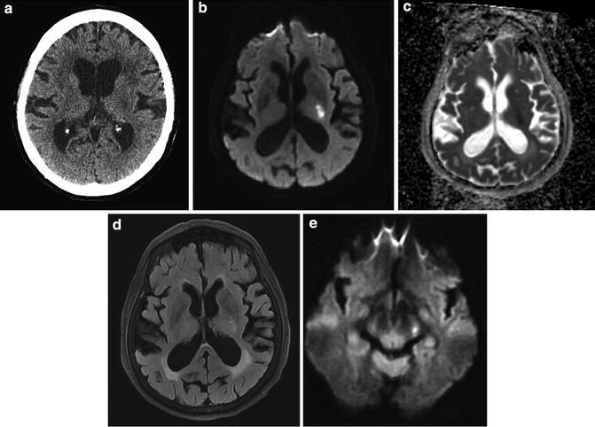
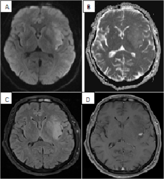
a DWI with intratumoral bleeding in a GBM patient without AT. b ...
Figure 1 from Guillain-Barré syndrome with optic neuritis and a focal ...
Reversible Splenial Lesion of the Corpus Callosum in Migrain... : The ...
Magnetic resonance imaging of brain reveals hypointense signals in ...
Neurocysticercosis - Neurologic Clinics
Magnetic Resonance Imaging Showing Left Occipital Lobe Lymphoma Before ...
A Pictorial Review on Reversible Splenial Lesions. - Abstract - Europe PMC
Case 34-2017 — A 76-Year-Old Man with Fever, Weight Loss, and Weakness ...
Chapter 13 – Neuroimaging in the Perioperative Neurocognitive Disorders ...
Parkinson's Brain Mri Vs Normal - ParkinsonsInfoClub.com
Neuroimaging in Postinfectious Demyelination and Nutritional Disorders ...
Axial brain MRI showing atrophy of the medial temporal lobes (A ...
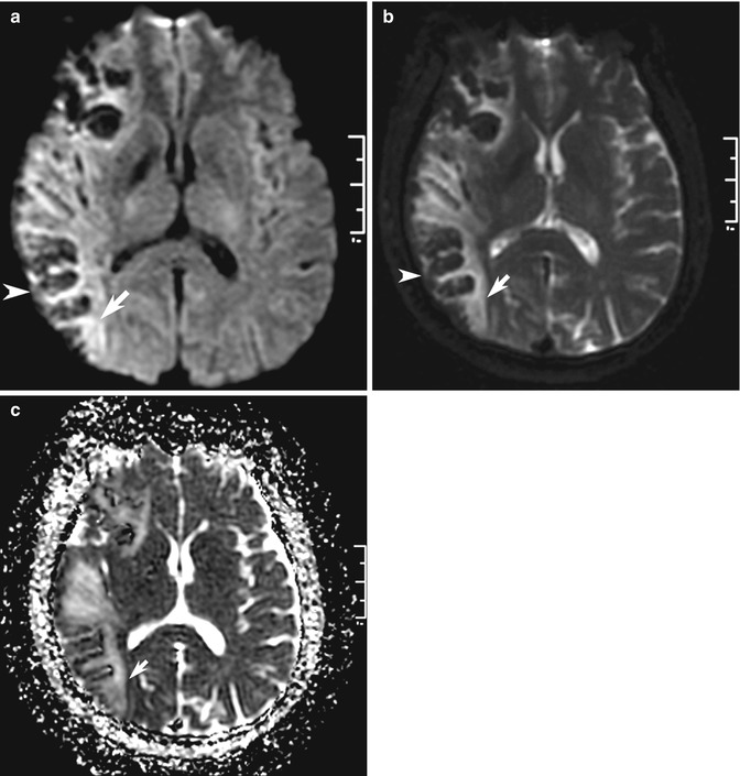
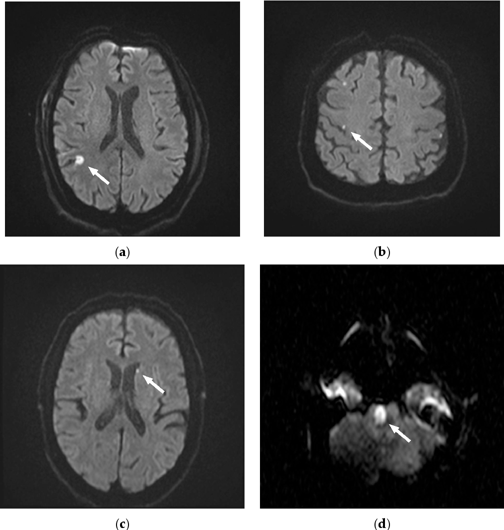
Examples of IALT localizations in the DWI sequence: A left anterior B ...
Diffusion MR Imaging: An Important Tool in the Assessment of Brain ...
Relationship between β-amyloid protein 1-42, thyroid hormone levels and ...
Magnetic resonance imaging of the brain. Scattered bilateral cerebral ...
Magnetic resonance imaging signs in human amyotrophic lateral sclerosis ...
75-year-old female patient with right side weakness. MRI was performed ...
Bupropion Overdose Presenting as Status Epilepticus in an Infant ...
Location of DAI lesions in the deep intra-axial structures. A and B ...
Diffusion weighted imaging shows regions of restricted diffusion in the ...
Marchiafava–Bignami disease-like lesions due to central nervous system ...
Frontiers | Distinct lesion features and underlying mechanisms in ...
Patterns of Ischemic Stroke: From Lacunar to Territorial to Multiple ...
Figure 1 from A splenial lesion with transiently reduced diffusion in ...
Brain | Radiology Key
Preoperative NCCT brain shows left frontal EDH, cerebral edema, and ...
Figure 2 from Frequency and Pattern of MRI Diffusion Restrictions after ...
Leukoaraiosis and Stroke | Stroke
Neuroimaging in Sickle Cell Disease: A Review - Mallon - 2020 - Journal ...
Prenatal Neurological Diagnosis: Challenges in Neuroimaging, Prognostic ...
Thrombolytic Therapy Is an Only Determinant Factor for Stroke Evolution ...
Anaplastic Astrocytoma Presenting as Ischemic Stroke: A Diagnostic Pitfall
Reversible MRI Changes in the Splenium Related to Recent Cessation of ...