Please enter url.
Login
Logout
Please enter url.
The MRI of Jahi McMath and Its Implications for the Global Ischemic ...
journals.sagepub.com
source
Comments
Figure 1 from The MRI of Jahi McMath and Its Implications for the ...
Trans-sulcal parafascicular approach to subcortical lesions - Journal ...
Neuroimaging of pediatric posterior fossa tumors including review of ...
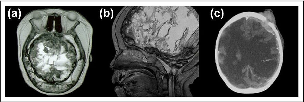
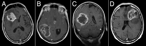
Pediatric multifocal glioblastoma multiforme with fulminant course ...
DVA associated with large basal ganglia hemorrhage. a Non-contrast head ...
Two different pediatric patients with BRAF V600E mutation gliomas ...
SciELO - Brasil - Multiple sporadic cerebral cavernous malformations ...
Tentorial meningioma with postoperative CT scan | Download Scientific ...
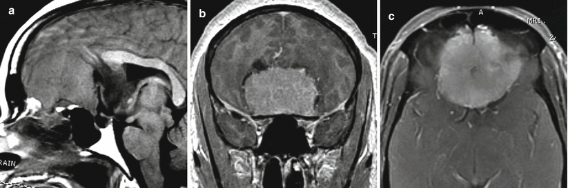
Meningioma of the Sellar and Parasellar Region | Neupsy Key
Primary Intracranial Atypical Teratoid/Rhabdoid Tumors of Infancy and ...
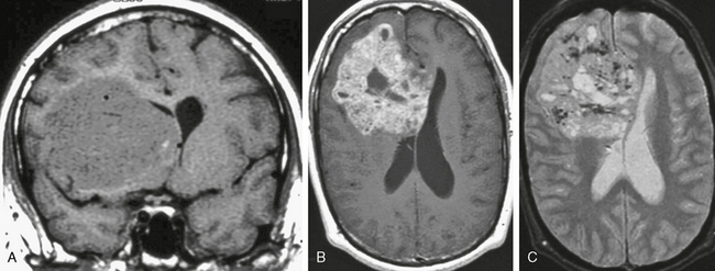
Figure 24 from Tumor or not tumor, that is the question. The ...
Figure 3 from Aneurysmal Bone Cyst of the Temporal Bone Presenting with ...
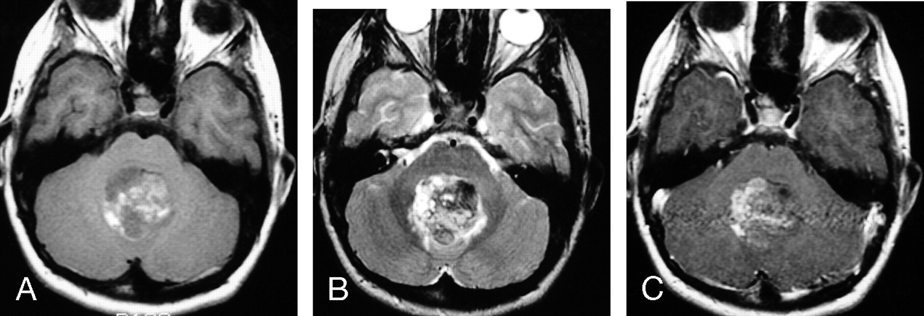
Magnetic resonance images before 7-fraction stereotactic radiosurgery ...
Post-operative MR images. a-c Sagittal non-contrast, axial ...
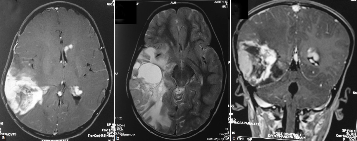
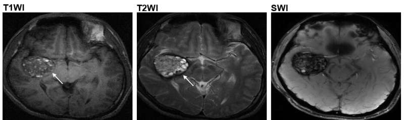
Neuroradiological representation of the lesions, T1WI showed a ...
(A) Axial T1W MRI with contrast showing radiological recurrence ...
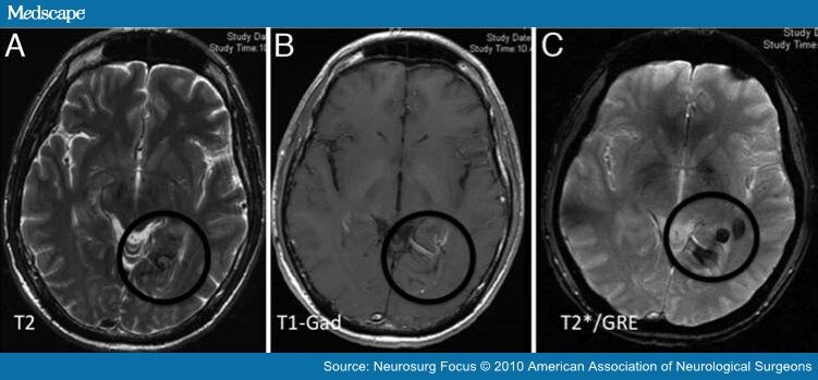
A-FLAIR, B-T1 + contrast, C-SWI. Lesion no. 3 located in the right ...
Infiltrative Gliomas | Neupsy Key
Pineal Region Masses | Radiology Key
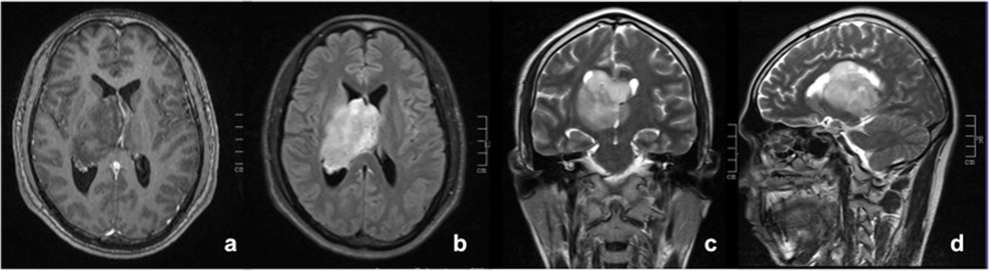
intraventricular neoplasms and lesions | pacs
Rapidly enlarging low-grade fibromyxoid sarcoma with intracranial ...
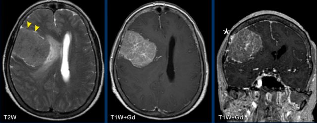
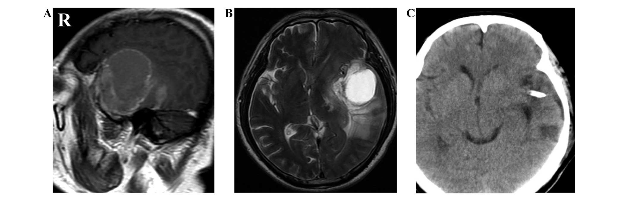
MRI at presentation. A) Axial diffusion-weighted image showing multiple ...
The Radiology Assistant : Enhancement Patterns in CNS disease
Magnetic resonance imaging (MRI) of the brain showing the cystic lesion ...
a–c Representative contrast-enhanced FSPGR MRI radiographs representing ...
Researchers discover a new cause for the cerebral cavernous malformation
IJERPH | Free Full-Text | Augmented Reality-Assisted Craniotomy for ...
Glioblastoma mimicking a cerebral contusion: A case report
Axial section brain MRI of our patient, showing a right temporal ...
Emerging Clinical Imaging Techniques for Cerebral Cavernous Malformations
Ventriculostomy Drain Mri Safe - Best Drain Photos Primagem.Org
Metastatic myxoid liposarcoma of the brain: a case report and review of ...
Congenital Melanocytic Nevi with Primary Cerebral Melanoma: A Rarity ...
Large Bifrontal Glioma | Operative Video Cases | The Neurosurgical Atlas
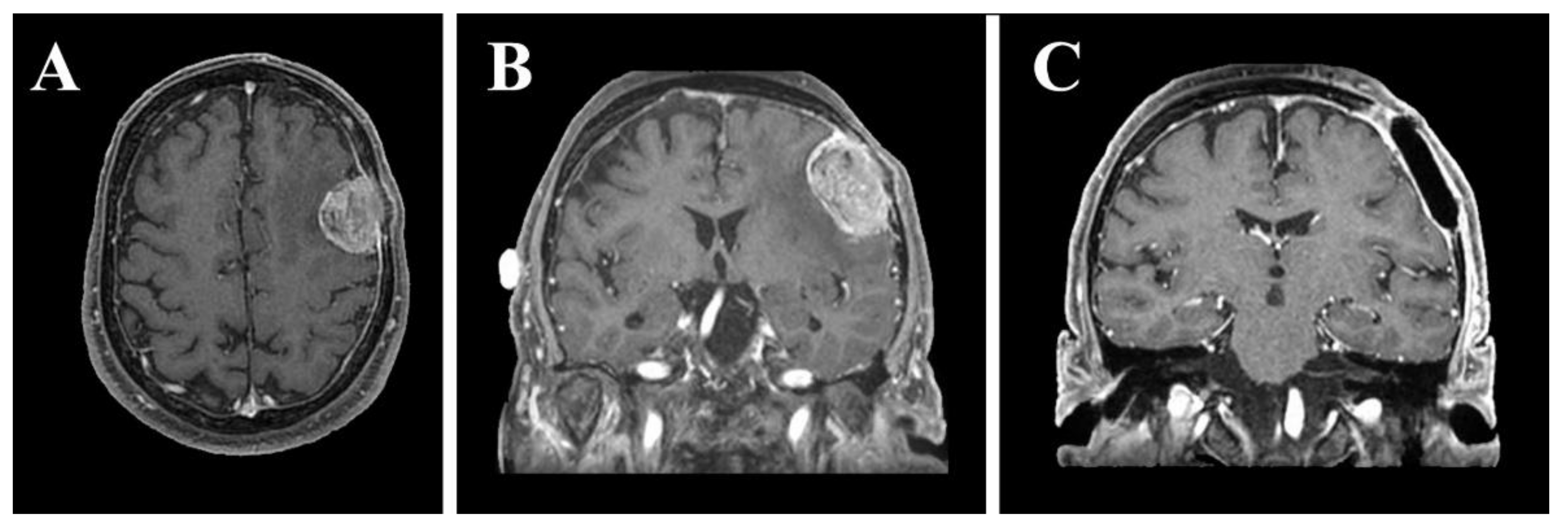
Malignant cystic meningioma of the falx cerebri (WHO grade III). a ...