Please enter url.
Login
Logout
Please enter url.
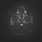
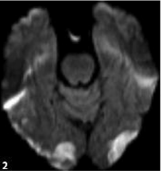
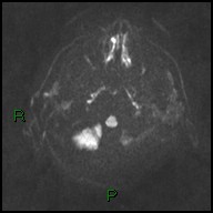
Axial T2 FSE (1) and MR-DWI (b value = 400) (2) images with ...
researchgate.net
source
Comments
Image | Radiopaedia.org
New method of assessment (TS/T method) | Download Scientific Diagram
A right incomplete medial infarction (arrows) of the middle and upper ...
36-year-old male with RRMS displays three lesions with better contrast ...
Coronal gadolinium-enhanced T1-weighted pituitary magnetic resonance ...
T1-weighted nonenhanced coronal MRI scan showing right-sided isointense ...
Preoperative Workup - Pituitary Adenomas - 78 Steps Health
Characteristics of lateral medullary infarction patients with hiccups ...
Image | Radiopaedia.org
Use of MRI in Acute Spinal Trauma | Radiology Key
Subependymoma | Image | Radiopaedia.org
Image | Radiopaedia.org
Image | Radiopaedia.org
Michel KALAMARIDES | Professor (Full) | MD PhD | Hôpital La Pitié ...
(PDF) Intervertebral Disk Degeneration in Dogs: Consequences, Diagnosis ...
Image | Radiopaedia.org
Anterior Cervical Fusion | Dr. Arvind Kulkarni
Dr Balaji Anvekar FRCR: Vascular territories of Brain stem and Infarct ...
Figure 2
» Things looking upCanadian Neuro-ophthalmology Group
Sella turcica magnetic resonance, coronal view, T1 weighted , with ...
Arachnoid cyst | Radiology Reference Article | Radiopaedia.org
Image | Radiopaedia.org
9 month old Turkish Van
Image | Radiopaedia.org
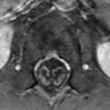
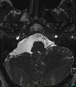
Solitary plasmacytoma involving C5 and C6 vertebrae with \'Mini brain ...
Dr Balaji Anvekar FRCR: Vascular territories of Brain stem and Infarct ...
Trigeminal neuralgia | Radiology Reference Article | Radiopaedia.org
Quantitative Computed Tomography (QCT) | UCSF Radiology
Pituitary Apoplexy: A Syndrome of Acute Visual Dysfunction - American ...
Enterovirus Rhombencephalomyelitis | Eurorad
Image | Radiopaedia.org
Sagittal and transverse T2 weighted MRI images of disc extrusion ...
(left) and (right) Standing x-rays done on D4 after the surgery showing ...
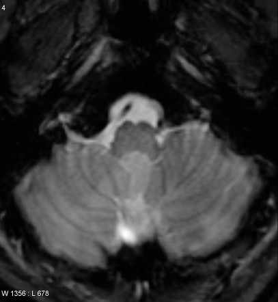
Post Gad: Right PICA Territory Infarct
Cor-T2-Brain-MRI
Cor-T2-Scoli-Abnormal-MRI
T1-MRI-Brain
T2-Stir-Cor-MRI
T2-TSE-Cor-MRI
MRI-Cor-T2-FSE-Right
Axial-T2-Brain-MRI
Cor-T2-Oblique-MRI
Normal-Brain-MRI-T2
MRI-Cor-OBL-T2-FS
T1-vs-T2-MRI-Spine
MRI-Brain-Sequences
Temporal-Lobe-MRI
Epilepsy-Protocol-MRI
T2-Cor-Pituitary-MRI
Sag-T1-Brain-MRI